

MSH6 Primary Antibody
Item Information
Catalog #
Size
Price
Description
This gene encodes a protein similar to the MutS protein. In E. coli, the MutS protein helps in the recognition of mismatched nucleotides, prior to their repair. A highly conserved region of approximately 150 aa, called the Walker-A adenine nucleotide binding motif, exists in MutS homologs. The encoded protein of this gene combines with MSH2 to form a mismatch recognition complex that functions as a bidirectional molecular switch that exchanges ADP and ATP as DNA mismatches are bound and dissociated. Mutations in this gene have been identified in individuals with hereditary nonpolyposis colon cancer (HNPCC) and endometrial cancer.Â
Product Overview
Entrez GenelD
2956
Aliases
GTBP; HSAP; HNPCC5
Clone#
5B11
Host / Isotype
Mouse / IgG1
Species Reactivity
Human
Immunogen
Purified recombinant fragment of human MSH6 expressed in E. Coli.
Formulation
Ascitic fluid containing 0.03% sodium azide.
Storage
Store at 4°C short term. Aliquot and store at -20°C long term. Avoid freeze/thaw cycles.
Product Applications
WB (Western Blot)
1/500 - 1/2000
ELISA
1/10000
References
1. J Biol Chem. 2009 Dec 11;284(50):34531-7.
2. J Biomed Sci. 2009 Oct 23;16:97.
2. J Biomed Sci. 2009 Oct 23;16:97.
Product Image
Western Blot

Figure 1: Western blot analysis using MSH6 mAb against human MSH6 (AA: 217-395) recombinant protein. (Expected MW is 45.5 kDa)
Western Blot
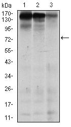
Figure 2: Western blot analysis using MSH6 mouse mAb against MCF-7 (1), HEK293 (2), and HCT116 (3) cell lysate.
Elisa

Black line: Control Antigen (100 ng); Purple line: Antigen(10ng); Blue line: Antigen (50 ng); Red line: Antigen (100 ng);
For Research Use Only. Not for use in diagnostic procedures.