

MSH2 Primary Antibody
Item Information
Catalog #
Size
Price
Description
MSH2 is a 100 kDa nuclear antigen and encodes a protein of 934 amino acids. The MSH2 gene is one of 4 known genes encoding proteins involved in the repair of mismatch nucleotides following DNA
replication or repair. Mutations in the MSH2 gene contribute to the development of sporadic
colorectal carcinoma. MSHS mutations are responsible for 50% of inherited non-polyposis colorectal
(HNPCC). The repair of mismatch DNA is essential to maintaining the integrity of genetic
information over time. An alteration of microsatellite repeats is the result of slippage owing to strand misalignment during DNA replication and is referred to as microsatellite instability (MSI).
These defects in DNA repair pathways have been related to human carcinogenesis. MSH-2 is involved in the initial cognition of mismatch nucleotides during the replication mismatch repair process.
Product Overview
Entrez GenelD
4436
Aliases
FCC1; COCA1; HNPCC; LCFS2
Clone#
1B3A8A8
Host / Isotype
Mouse / IgG1
Species Reactivity
Human, Monkey
Immunogen
Purified recombinant fragment of human MSH2 expressed in E. Coli.
Formulation
Ascitic fluid containing 0.03% sodium azide.
Storage
Store at 4°C short term. Aliquot and store at -20°C long term. Avoid freeze/thaw cycles.
Product Applications
WB (Western Blot)
1/500 - 1/2000
IHC_P(Immunohistochemistry)
1/200 - 1/1000
ICC (Immunocytochemistry)
1/200 - 1/1000
ELISA
1/10000
References
1. Papadopoulos, N. 1994. Science 263: 1625-1629.
2. Palombo, F. 1994. Nature 367:417-418.
2. Palombo, F. 1994. Nature 367:417-418.
Product Image
Western Blot
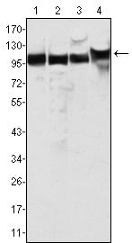
Figure 1: Western blot analysis using MSH2 mouse mAb against Hela (1), A549 (2), A431 (3) and HEK293 (4) cell lysate.
Immunohistochemical analysis

Figure 2: Immunohistochemical analysis of paraffin-embedded human breast cancer (left) and lung cancer (right) tissues, showing nuclear localization using MSH2 mouse mAb with DAB staining.
Immunofluorescence analysis

Figure 3: Confocal Immunofluorescence analysis of Hela cells using MSH2 mouse mAb (green), showing nuclear localization. Red: Actin filaments have been labeled with Alexa Fluor-555 phalloidin.
For Research Use Only. Not for use in diagnostic procedures.