

MRPL42 Primary Antibody
Item Information
Catalog #
Size
Price
Description
Mammalian mitochondrial ribosomal proteins are encoded by nuclear genes and help in protein synthesis within the mitochondrion. Mitochondrial ribosomes (mitoribosomes) consist of a small 28S subunit and a large 39S subunit. They have an estimated 75% protein to rRNA composition compared to prokaryotic ribosomes, where this ratio is reversed. Another difference between mammalian mitoribosomes and prokaryotic ribosomes is that the latter contain a 5S rRNA. Among different species, the proteins comprising the mitoribosome differ greatly in sequence, and sometimes in biochemical properties, which prevents easy recognition by sequence homology. This gene encodes a protein identified as belonging to both the 28S and the 39S subunits. Alternative splicing results in multiple transcript variants. Pseudogenes corresponding to this gene are found on chromosomes 4q, 6p, 6q, 7p, and 15q.
Product Overview
Entrez GenelD
28977
Aliases
L31MT; L42MT; S32MT; MRPL31; MRPS32; PTD007; RPML31; HSPC204; MRP-L31; MRP-L42; MRP-S32
Clone#
3H6H2
Host / Isotype
Mouse / IgG1
Species Reactivity
Human
Immunogen
Purified recombinant fragment of human MRPL42 (AA: 10-142) expressed in E. Coli.
Formulation
Purified antibody in PBS with 0.05% sodium azide
Storage
Store at 4°C short term. Aliquot and store at -20°C long term. Avoid freeze/thaw cycles.
Product Applications
WB (Western Blot)
1/500 - 1/2000
IHC_P(Immunohistochemistry)
1/200 - 1/1000
FCM (Flow Cytometry)
1/200 - 1/400
ELISA
1/10000
References
1. Genomics. 2003 May;81(5):468-80.
2. J Biol Chem. 2001 Nov 23;276(47):43958-69.
2. J Biol Chem. 2001 Nov 23;276(47):43958-69.
Product Image
Western Blot

Figure 1: Western blot analysis using MRPL42 mAb against human MRPL42 recombinant protein. (Expected MW is 41.2 kDa)
Western Blot

Figure 2: Western blot analysis using MRPL42 mAb against HEK293 (1) and MRPL42 (AA: 10-142)-hIgGFc transfected HEK293 (2) cell lysate.
Western Blot

Figure 3: Western blot analysis using MRPL42 mouse mAb against HL7702 (1), SMMC-7721 (2), HEK293 (3) , HeLa (4) and Raji (5) cell lysate.
Flow cytometric

Figure 4: Flow cytometric analysis of HepG2 cells using MRPL42 mouse mAb (green) and negative control (purple).
Immunohistochemical analysis
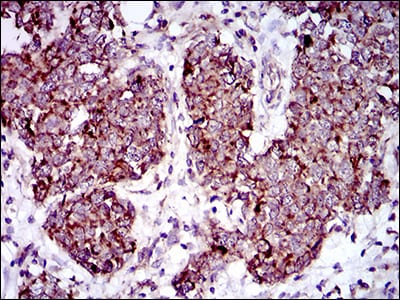
Figure 5: Immunohistochemical analysis of paraffin-embedded breast cancer tissues using MRPL42 mouse mAb with DAB staining.
Immunohistochemical analysis
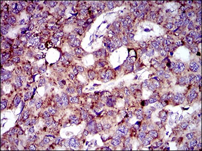
Figure 6: Immunohistochemical analysis of paraffin-embedded esophageal cancer tissues using MRPL42 mouse mAb with DAB staining.
Elisa

Black line: Control Antigen (100 ng); Purple line: Antigen(10ng); Blue line: Antigen (50 ng); Red line: Antigen (100 ng);
For Research Use Only. Not for use in diagnostic procedures.