

MRE11 Primary Antibody
Item Information
Catalog #
Size
Price
Description
This gene encodes a nuclear protein involved in homologous recombination, telomere length maintenance, and DNA double-strand break repair. By itself, the protein has 3' to 5' exonuclease activity and endonuclease activity. The protein forms a complex with the RAD50 homolog; this complex is required for nonhomologous joining of DNA ends and possesses increased single-stranded DNA endonuclease and 3' to 5' exonuclease activities. In conjunction with a DNA ligase, this protein promotes the joining of noncomplementary ends in vitro using short homologies near the ends of the DNA fragments. This gene has a pseudogene on chromosome 3. Alternative splicing of this gene results in two transcript variants encoding different isoforms.
Product Overview
Entrez GenelD
4361
Aliases
ATLD; HNGS1; MRE11A; MRE11B
Clone#
7C8A9
Host / Isotype
Mouse / Mouse IgG2b
Species Reactivity
Human, Mouse, Monkey, Rat
Immunogen
Purified recombinant fragment of human MRE11 (AA: 182-582) expressed in E. Coli.
Formulation
Purified antibody in PBS with 0.05% sodium azide
Storage
Store at 4°C short term. Aliquot and store at -20°C long term. Avoid freeze/thaw cycles.
Product Applications
WB (Western Blot)
1/500 - 1/2000
IHC_P(Immunohistochemistry)
1/200 - 1/1000
FCM (Flow Cytometry)
1/200 - 1/400
ELISA
1/10000
References
Cancer Lett. 2021 Aug 28;514:1-11.
Diagn Pathol. 2019 Jun 21;14(1):60.
Diagn Pathol. 2019 Jun 21;14(1):60.
Product Image
Elisa
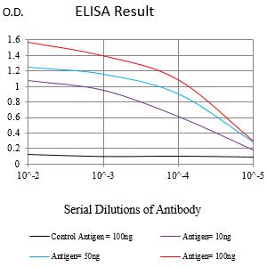
Figure 1:Black line: Control Antigen (100 ng);Purple line: Antigen (10ng); Blue line: Antigen (50 ng); Red line:Antigen (100 ng)
Western Blot

Figure 2:Western blot analysis using MRE11 mouse mAb against Hela (1), A431 (2), MCF-7 (3), Jurkat (4), HepG2 (5), K562 (6), COS-7 (7), PC-12 (8) and NIH/3T3 (9) cell lysate.
Immunofluorescence analysis

Figure 3:Flow cytometric analysis of Hela cells using MRE11 mouse mAb (green) and negative control (red).
Immunofluorescence analysis

Figure 4:Flow cytometric analysis of K562 cells using MRE11 mouse mAb (green) and negative control (red).
Immunohistochemical analysis

Figure 5:Immunohistochemical analysis of paraffin-embedded ovarian cancer tissues using MRE11 mouse mAb with DAB staining.
Immunohistochemical analysis

Figure 6:Immunohistochemical analysis of paraffin-embedded rectal cancer tissues using MRE11 mouse mAb with DAB staining.
Immunohistochemical analysis

Figure 7:Immunohistochemical analysis of paraffin-embedded Mouse testis tissues using MRE11 mouse mAb with DAB staining.
Immunohistochemical analysis
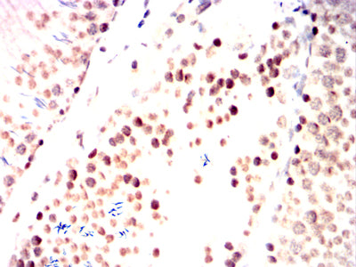
Figure 8:Immunohistochemical analysis of paraffin-embedded Rat testis tissues using MRE11 mouse mAb with DAB staining.
Immunohistochemical analysis

Figure 9:Immunohistochemical analysis of paraffin-embedded Rabbit testis tissues using MRE11 mouse mAb with DAB staining.
For Research Use Only. Not for use in diagnostic procedures.