

mouse Lplunc1 Primary Antibody
Item Information
Catalog #
Size
Price
Description
Palate, lung, and nasal epithelium clone (Plunc, now renamed Splunc1) is a small secreted protein expressed in the oropharynx and upper airways of humans, mice, rats, and cows. This protein is structurally homologous to known mediators of host defense against gram-negative bacteria.
Product Overview
Entrez GenelD
228801
Aliases
Bpifb1; RP23-154J12.1
Clone#
4H2
Host / Isotype
Mouse / IgG1
Species Reactivity
Human
Immunogen
Purified recombinant fragment of mouse Lplunc1 expressed in E. Coli.
Formulation
Purified antibody in PBS with 0.05% sodium azide
Storage
Store at 4°C short term. Aliquot and store at -20°C long term. Avoid freeze/thaw cycles.
Product Applications
WB (Western Blot)
1/500 - 1/2000
IHC_P(Immunohistochemistry)
1/200 - 1/1000
ICC (Immunocytochemistry)
1/200 - 1/1000
FCM (Flow Cytometry)
1/200 - 1/400
ELISA
1/10000
References
1. Briand F, et al. Clin Transl Sci, 2011 Dec.
2. Tang T, et al. Nat Biotechnol, 2010 Jul
2. Tang T, et al. Nat Biotechnol, 2010 Jul
Product Image
Western Blot

Figure 1: Western blot analysis using Lplunc1 mAb against mouse Lplunc1 (AA: 248-475) recombinant protein. (Expected MW is 50.8 kDa)
Immunohistochemical analysis
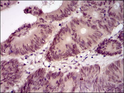
Figure 2: Immunohistochemical analysis of paraffin-embedded colon cancer tissues using Lplunc1 mouse mAb with DAB staining.
Immunohistochemical analysis

Figure 2: Immunohistochemical analysis of paraffin-embedded muscle tissues using Lplunc1 mouse mAb with DAB staining.
Flow cytometric

Figure 4: Flow cytometric analysis of MCF-7 cells using Lplunc1 mouse mAb (green) and negative control (red).
Immunofluorescence analysis

Figure 5: Immunofluorescence analysis of HeLa cells using Lplunc1 mouse mAb (green). Blue: DRAQ5 fluorescent DNA dye. Red: Actin filaments have been labeled with Alexa Fluor-555 phalloidin.
Elisa

Black line: Control Antigen (100 ng); Purple line: Antigen(10ng); Blue line: Antigen (50 ng); Red line: Antigen (100 ng);
For Research Use Only. Not for use in diagnostic procedures.