
MMP9 Primary Antibody
Item Information
Catalog #
Size
Price
Description
Proteins of the matrix metalloproteinase (MMP) family are involved in the breakdown of extracellular matrix in normal physiological processes, such as embryonic development, reproduction, and tissue remodeling, as well as in disease processes, such as arthritis and metastasis. Most MMP's are secreted as inactive proproteins which are activated when cleaved by extracellular proteinases. The enzyme encoded by this gene degrades type IV and V collagens. Studies in rhesus monkeys suggest that the enzyme is involved in IL-8-induced mobilization of hematopoietic progenitor cells from bone marrow, and murine studies suggest a role in tumor-associated tissue remodeling.
Product Overview
Entrez GenelD
4318
Aliases
GELB; CLG4B; MMP-9; MANDP2
Clone#
5G3
Host / Isotype
Mouse / IgG2a
Species Reactivity
Human, Mouse
Immunogen
Purified recombinant fragment of human MMP9 expressed in E. Coli.
Formulation
Ascitic fluid containing 0.03% sodium azide.
Storage
Store at 4°C short term. Aliquot and store at -20°C long term. Avoid freeze/thaw cycles.
Product Applications
WB (Western Blot)
1/500 - 1/2000
IHC_P(Immunohistochemistry)
1/200 - 1/1000
ICC (Immunocytochemistry)
1/200 - 1/1000
FCM (Flow Cytometry)
1/200 - 1/400
ELISA
1/10000
References
1. IUBMB Life. 2009 Dec;61(12):1143-52.
2. J Biol Regul Homeost Agents. 2009 Oct-Dec;23(4):259-67.
2. J Biol Regul Homeost Agents. 2009 Oct-Dec;23(4):259-67.
Product Image
Western Blot

Figure 1: Western blot analysis using MMP9 mAb against human MMP9 (AA: 238-465) recombinant protein. (Expected MW is 50.6 kDa)
Western Blot
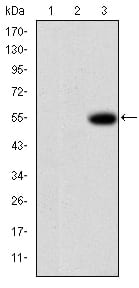
Figure 2: Western blot analysis using MMP9 mAb against HEK293 (1), MMP7-hIgGFc transfected HEK293 (2) cell lysate and MMP9-hIgGFc transfected HEK293 (3) cell lysate.
Immunohistochemical analysis

Figure 3: Immunohistochemical analysis of paraffin-embedded brain tissues using MMP9 mouse mAb with DAB staining.
Immunofluorescence analysis
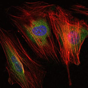
Figure 4: Immunofluorescence analysis of NIH/3T3 cells using MMP9 mouse mAb (green). Blue: DRAQ5 fluorescent DNA dye. Red: Actin filaments have been labeled with Alexa Fluor-555 phalloidin.
Flow cytometric
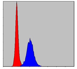
Figure 5: Flow cytometric analysis of Hela cells using MMP9 mouse mAb (blue) and negative control (red).
Elisa
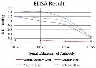
Red: Control Antigen (100ng); Purple: Antigen (10ng); Green: Antigen (50ng); Blue: Antigen (100ng);
For Research Use Only. Not for use in diagnostic procedures.