
MMP1 Primary Antibody
Item Information
Catalog #
Size
Price
Description
Proteins of the matrix metalloproteinase (MMP) family are involved in the breakdown of extracellular matrix in normal physiological processes, such as embryonic development, reproduction, and tissue remodeling, as well as in disease processes, such as arthritis and metastasis. Most MMP's are secreted as inactive proproteins which are activated when cleaved by extracellular proteinases. This gene encodes a secreted enzyme which breaks down the interstitial collagens, types I, II, and III. The gene is part of a cluster of MMP genes which localize to chromosome 11q22.3. Alternative splicing results in multiple transcript variants.
Product Overview
Entrez GenelD
4312
Aliases
CLG; CLGN
Clone#
6A5
Host / Isotype
Mouse / IgG1
Species Reactivity
Human
Immunogen
Purified recombinant fragment of human MMP1 expressed in E. Coli.
Formulation
Ascitic fluid containing 0.03% sodium azide.
Storage
Store at 4°C short term. Aliquot and store at -20°C long term. Avoid freeze/thaw cycles.
Product Applications
WB (Western Blot)
1/500 - 1/2000
IHC_P(Immunohistochemistry)
1/200 - 1/1000
ICC (Immunocytochemistry)
1/200 - 1/1000
FCM (Flow Cytometry)
1/200 - 1/400
ELISA
1/10000
References
1. Arthritis Res Ther. 2009;11(6):R169.
2. FEMS Microbiol Lett. 2009 Oct;299(2):214-22.
2. FEMS Microbiol Lett. 2009 Oct;299(2):214-22.
Product Image
Western Blot
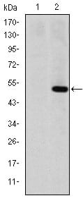
Figure 1: Western blot analysis using MMP1 mAb against HEK293 (1) and MMP1(AA: 24-213)-hIgGFc transfected HEK293 (2) cell lysate.
Immunohistochemical analysis

Figure 2: Immunohistochemical analysis of paraffin-embedded human cervical cancer tissues (left) and human kidney cancer tissues (right) using MMP1 mouse mAb with DAB staining.
Flow cytometric

Figure 3: Flow cytometric analysis of Hela cells using MMP1 mouse mAb (green) and negative control (purple).
Immunofluorescence analysis

Figure 4: Immunofluorescence analysis of Hela cells using MMP1 mouse mAb (green). Blue: DRAQ5 fluorescent DNA dye. Red: Actin filaments have been labeled with Alexa Fluor-555 phalloidin.
Elisa

Red: Control Antigen (100ng); Purple: Antigen (10ng); Green: Antigen (50ng); Blue: Antigen (100ng);
For Research Use Only. Not for use in diagnostic procedures.