

MLXIPL Primary Antibody
Item Information
Catalog #
Size
Price
Description
This gene encodes a basic helix-loop-helix leucine zipper transcription factor of the Myc/Max/Mad superfamily. This protein forms a heterodimeric complex and binds and activates, in a glucose-dependent manner, carbohydrate response element (ChoRE) motifs in the promoters of triglyceride synthesis genes. The gene is deleted in Williams-Beuren syndrome, a multisystem developmental disorder caused by the deletion of contiguous genes at chromosome 7q11.23.
Product Overview
Entrez GenelD
51085
Aliases
MIO; CHREBP; MONDOB; WBSCR14; WS-bHLH; bHLHd14
Clone#
5D12D1
Host / Isotype
Mouse / IgG1
Species Reactivity
Human
Immunogen
Purified recombinant fragment of human MLXIPL (AA: 18-143) expressed in E. Coli.
Formulation
Ascitic fluid containing 0.03% sodium azide.
Storage
Store at 4°C short term. Aliquot and store at -20°C long term. Avoid freeze/thaw cycles.
Product Applications
WB (Western Blot)
1/500 - 1/2000
ICC (Immunocytochemistry)
1/200 - 1/1000
ELISA
1/10000
References
1. Diabetes. 2012 Mar;61(3):574-85.
2. Biochim Biophys Acta. 2011 Dec;1811(12):1194-200.
2. Biochim Biophys Acta. 2011 Dec;1811(12):1194-200.
Product Image
Western Blot
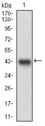
Figure 1: Western blot analysis using MLXIPL mAb against human MLXIPL recombinant protein. (Expected MW is 41 kDa)
Western Blot

Figure 2: Western blot analysis using MLXIPL mAb against HEK293 (1) and MLXIPL (AA: 18-143)-hIgGFc transfected HEK293 (2) cell lysate.
Immunofluorescence analysis

Figure 3: Immunofluorescence analysis of Hela cells using MLXIPL mouse mAb (green). Blue: DRAQ5 fluorescent DNA dye. Red: Actin filaments have been labeled with Alexa Fluor-555 phalloidin.
Elisa
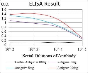
Black line: Control Antigen (100 ng); Purple line: Antigen(10ng); Blue line: Antigen (50 ng); Red line: Antigen (100 ng);
For Research Use Only. Not for use in diagnostic procedures.