
MIB1 Primary Antibody
Item Information
Catalog #
Size
Price
Description
This gene encodes a protein containing multiple ankyrin repeats and RING finger domains that functions as an E3 ubiquitin ligase. The encoded protein positively regulates Notch signaling by ubiquitinating the Notch receptors, thereby facilitating their endocytosis. This protein may also promote the ubiquitination and degradation of death-associated protein kinase 1 (DAPK1).
Product Overview
Entrez GenelD
57534
Aliases
MIB; DIP1; ZZZ6; DIP-1; LVNC7; ZZANK2
Clone#
2A7B1
Host / Isotype
Mouse / IgG1
Species Reactivity
Human, Monkey
Immunogen
Purified recombinant fragment of human MIB1 (AA: 6-221) expressed in E. Coli.
Formulation
Purified antibody in PBS with 0.05% sodium azide
Storage
Store at 4°C short term. Aliquot and store at -20°C long term. Avoid freeze/thaw cycles.
Product Applications
WB (Western Blot)
1/500 - 1/2000
FCM (Flow Cytometry)
1/200 - 1/400
ELISA
1/10000
References
1.J Cell Sci. 2015 May 1;128(9):1674-82.
2.Cell Res. 2012 Mar;22(3):603-6.
2.Cell Res. 2012 Mar;22(3):603-6.
Product Image
Elisa
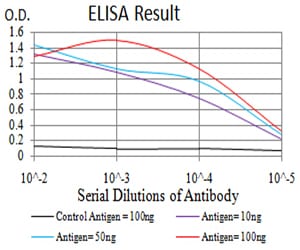
Figure 1: Black line: Control Antigen (100 ng);Purple line: Antigen (10ng); Blue line: Antigen (50 ng); Red line:Antigen (100 ng)
Western Blot
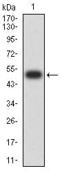
Figure 2:Western blot analysis using MIB1 mAb against human MIB1 (AA: 6-221) recombinant protein. (Expected MW is 50.1 kDa)
Western Blot
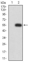
Figure 3:Western blot analysis using MIB1 mAb against HEK293 (1) and MIB1 (AA: 6-221)-hIgGFc transfected HEK293 (2) cell lysate.
Western Blot
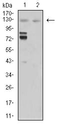
Figure 4:Western blot analysis using MIB1 mouse mAb against Hela (1) and COS7 (2) cell lysate.
Flow cytometric
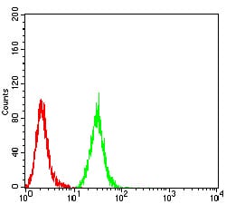
Figure 5:Flow cytometric analysis of Hela cells using MIB1 mouse mAb (green) and negative control (red).
For Research Use Only. Not for use in diagnostic procedures.