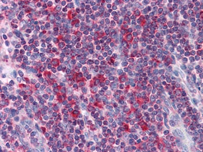
Metadherin Primary Antibody

Item Information
Catalog #
Size
Price
Description

Metadherin (Metastasis adhesion protein), also known as MTDH, LYsine-RIch CEACAM1 co-isolated (LYRIC), is a novel protein that localizes with the tight junction proteins ZO-1 and occludin in polarized epithelial cells. At the tight junction, it acts not as a structural component, but is rather recruited during the maturation of the tight junction complex. Metadherin is overexpressed in breast cancer tissue and breast tumor xenografts, while much lower levels are expressed in normal breast tissue. Metadherin binds to lung vasculature, one of the four common sites of breast cancer metastasis, through a C-terminal segment in the extracellular domain; blocking this lung-homing domain with antibodies or inhibiting metadherin with siRNA has been reported to inhibit breast cancer metastasis.

Product Overview
Entrez GenelD
92140
Aliases
3D3; AEG1; LYRIC; MTDH
Clone#
2F11C3
Host / Isotype
Mouse / IgG1
Species Reactivity
Human
Immunogen
Purified recombinant fragment of human Metadherin expressed in E. Coli.
Formulation
Ascitic fluid containing 0.03% sodium azide.
Storage
4°C; -20°C for long term storage
Product Applications
WB (Western Blot)
1/500 - 1/2000
IHC_P(Immunohistochemistry)
1/200 - 1/1000
FCM (Flow Cytometry)
1/200 - 1/400
ELISA
1/10000
References
1. Cancer Cell. 2004 Apr;5(4):365-74.
2. Exp Cell Res. 2004 Oct 15;300(1):134-48.
3. Cancer Cell. 2009 Jan 6;15(1):9-20.
Product Image
Western Blot
Figure 1: Western blot analysis using Metadherin mouse mAb against K562 (1), SKBR-3 (2), T47D (3), Hela (4) and MCF-7 (5) cell lysate.
Immunohistochemical analysis
Figure 2: Immunohistochemical analysis of paraffin-embedded human Liver tissues using Metadherin mouse mAb
Flow cytometric
Figure 3: Flow cytometric analysis of Hela cells using Metadherin mouse mAb (green) and negative control (purple).
For Research Use Only. Not for use in diagnostic procedures.