

MET Primary Antibody
Item Information
Catalog #
Size
Price
Description
This gene encodes a member of the receptor tyrosine kinase family of proteins and the product of the proto-oncogene MET. The encoded preproprotein is proteolytically processed to generate alpha and beta subunits that are linked via disulfide bonds to form the mature receptor. Further processing of the beta subunit results in the formation of the M10 peptide, which has been shown to reduce lung fibrosis. Binding of its ligand, hepatocyte growth factor, induces dimerization and activation of the receptor, which plays a role in cellular survival, embryogenesis, and cellular migration and invasion. Mutations in this gene are associated with papillary renal cell carcinoma, hepatocellular carcinoma, and various head and neck cancers. Amplification and overexpression of this gene are also associated with multiple human cancers.
Product Overview
Entrez GenelD
4233
Aliases
HGFR; AUTS9; RCCP2; c-Met; DFNB97
Clone#
5E9C4
Host / Isotype
Mouse / IgG1
Species Reactivity
Human, Monkey
Immunogen
Purified recombinant fragment of human MET (AA: 743-932) expressed in E. Coli.
Formulation
Purified antibody in PBS with 0.05% sodium azide
Storage
4°C; -20°C for long term storage
Product Applications
WB (Western Blot)
1/500 - 1/2000
IHC_P(Immunohistochemistry)
1/200 - 1/1000
ICC (Immunocytochemistry)
1/200 - 1/1000
FCM (Flow Cytometry)
1/200 - 1/400
ELISA
1/10000
References
1.Oncotarget. 2015 Jun 30;6(18):16215-26.
2.Br J Cancer. 2015 Feb 3;112(3):429-37.
2.Br J Cancer. 2015 Feb 3;112(3):429-37.
Product Image
Elisa
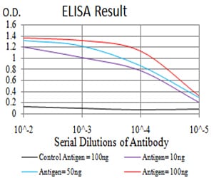
Figure 1: Black line: Control Antigen (100 ng);Purple line: Antigen (10ng); Blue line: Antigen (50 ng); Red line:Antigen (100 ng)
Western Blot
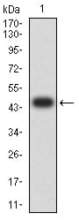
Figure 2:Western blot analysis using MET mAb against human MET (AA: 743-932) recombinant protein. (Expected MW is 51 kDa)
Western Blot
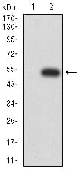
Figure 3:Western blot analysis using MET mAb against HEK293 (1) and MET (AA: 743-932)-hIgGFc transfected HEK293 (2) cell lysate.
Western Blot
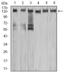
Figure 4:Western blot analysis using MET mouse mAb against A549 (1), COS7 (2), Hela (3), HEK293 (4), HepG2 (5), and A431 (6) cell lysate.
Immunofluorescence analysis
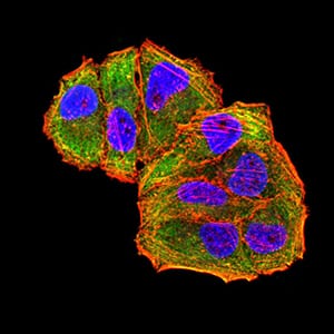
Figure 5:Immunofluorescence analysis of Hela cells using MET mouse mAb (green). Blue: DRAQ5 fluorescent DNA dye. Red: Actin filaments have been labeled with Alexa Fluor- 555 phalloidin. Secondary antibody from Fisher (Cat#: 35503)
Flow cytometric
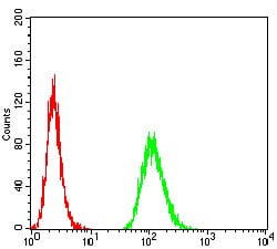
Figure 6:Flow cytometric analysis of MCF-7 cells using MET mouse mAb (green) and negative control (red).
Immunohistochemical analysis
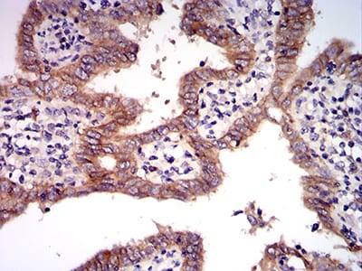
Figure 7:Immunohistochemical analysis of paraffin-embedded endometrial cancer tissues using MET mouse mAb with DAB staining.
For Research Use Only. Not for use in diagnostic procedures.