
MEN1 Primary Antibody
Item Information
Catalog #
Size
Price
Description
This gene encodes menin, a putative tumor suppressor associated with a syndrome known as multiple endocrine neoplasia type 1. In vitro studies have shown menin is localized to the nucleus, possesses two functional nuclear localization signals, and inhibits transcriptional activation by JunD, however, the function of this protein is not known. Two messages have been detected on northern blots but the larger message has not been characterized. Alternative splicing results in multiple transcript variants.
Product Overview
Entrez GenelD
4221
Aliases
MEAI; SCG2
Clone#
7D3E10
Host / Isotype
Mouse / IgG1
Species Reactivity
Human
Immunogen
Purified recombinant fragment of human MEN1 (AA: 392-554) expressed in E. Coli.
Formulation
Purified antibody from tissue culture in PBS with 0.05% sodium azide
Storage
Store at 4°C short term. Aliquot and store at -20°C long term. Avoid freeze/thaw cycles.
Product Applications
WB (Western Blot)
1/500 - 1/2000
IHC_P(Immunohistochemistry)
1/200 - 1/1000
FCM (Flow Cytometry)
1/200 - 1/400
ELISA
1/10000
References
1. Clinics (Sao Paulo). 2012;67 Suppl 1:49-56.
2. World J Surg Oncol. 2011 Jan 25;9:6.
2. World J Surg Oncol. 2011 Jan 25;9:6.
Product Image
Western Blot
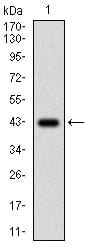
Figure 1: Western blot analysis using MEN1 mAb against human MEN1 (AA: 392-554) recombinant protein. (Expected MW is 43.3 kDa)
Western Blot

Figure 2: Western blot analysis using MEN1 mAb against HEK293 (1) and MEN1 (AA: 392-554)-hIgGFc transfected HEK293 (2) cell lysate.
Flow cytometric
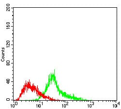
Figure 3: Flow cytometric analysis of Hela cells using MEN1 mouse mAb (green) and negative control (red).
Immunohistochemical analysis
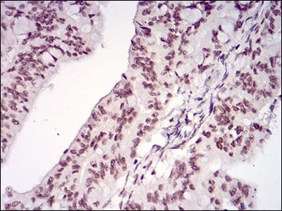
Figure 4: Immunohistochemical analysis of paraffin-embedded rectum cancer tissues using MEN1 mouse mAb with DAB staining.
Elisa
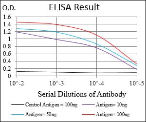
Black line: Control Antigen (100 ng); Purple line: Antigen(10ng); Blue line: Antigen (50 ng); Red line: Antigen (100 ng);
For Research Use Only. Not for use in diagnostic procedures.