

MEF2A Primary Antibody
Item Information
Catalog #
Size
Price
Description
The protein encoded by this gene is a DNA-binding transcription factor that activates many muscle-specific, growth factor-induced, and stress-induced genes. The encoded protein can act as a homodimer or as a heterodimer and is involved in several cellular processes, including muscle development, neuronal differentiation, cell growth control, and apoptosis. Defects in this gene could be a cause of autosomal dominant coronary artery disease 1 with myocardial infarction (ADCAD1). Several transcript variants encoding different isoforms have been found for this gene.
Product Overview
Entrez GenelD
4205
Aliases
mef2; ADCAD1; RSRFC4; RSRFC9
Clone#
6B6F8
Host / Isotype
Mouse / IgG1
Species Reactivity
Human
Immunogen
Purified recombinant fragment of human MEF2A (AA: 391-497) expressed in E. Coli.
Formulation
Purified antibody in PBS with 0.05% sodium azide
Storage
Store at 4°C short term. Aliquot and store at -20°C long term. Avoid freeze/thaw cycles.
Product Applications
WB (Western Blot)
1/500 - 1/2000
ICC (Immunocytochemistry)
1/200 - 1/1000
FCM (Flow Cytometry)
1/200 - 1/400
ELISA
1/10000
References
1. Cell Biochem Funct. 2012 Mar;30(2):108-13.
2. Circ Cardiovasc Genet. 2009 Apr;2(2):165-72.
2. Circ Cardiovasc Genet. 2009 Apr;2(2):165-72.
Product Image
Western Blot
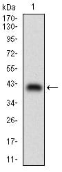
Figure 1: Western blot analysis using MEF2A mAb against human MEF2A (AA: 391-497) recombinant protein. (Expected MW is 38 kDa)
Western Blot

Figure 2: Western blot analysis using MEF2A mAb against HEK293 (1) and MEF2A (AA: 391-497)-hIgGFc transfected HEK293 (2) cell lysate.
Immunofluorescence analysis
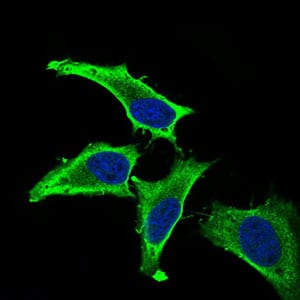
Figure 3: Immunofluorescence analysis of Hela cells using MEF2A mouse mAb (green). Blue: DRAQ5 fluorescent DNA dye. Secondary antibody from Fisher (Cat#: 35503)
Flow cytometric
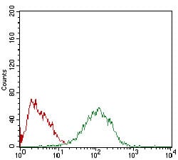
Figure 4: Flow cytometric analysis of HepG2 cells using MEF2A mouse mAb (green) and negative control (red).
Elisa

Black line: Control Antigen (100 ng); Purple line: Antigen(10ng); Blue line: Antigen (50 ng); Red line: Antigen (100 ng);
For Research Use Only. Not for use in diagnostic procedures.