

MDM4 Primary Antibody
Item Information
Catalog #
Size
Price
Description
MDM4, encodes a 490-amino acid protein containing a RING finger domain and a putative nuclear localization signal. The MDM4 putative nuclear localization signal, which all Mdm proteins contain, is located in the C-terminal region of the protein. The mRNA is expressed at a high level in thymus and at lower levels in all other tissues tested. MDM4 protein produced by in vitro translation interacts with p53 via a binding domain located in the N-terminal region of the MDM4 protein. MDM4 shows significant structural similarity to p53-binding protein MDM2.
Product Overview
Entrez GenelD
4194
Aliases
HDMX; MDMX; MRP1; MDM4
Clone#
2D10F4
Host / Isotype
Mouse / IgG1
Species Reactivity
Human
Immunogen
Purified recombinant fragment of human MDM4 expressed in E. Coli.
Formulation
Ascitic fluid containing 0.03% sodium azide.
Storage
Store at 4°C short term. Aliquot and store at -20°C long term. Avoid freeze/thaw cycles.
Product Applications
WB (Western Blot)
1/500 - 1/2000
IHC_P(Immunohistochemistry)
1/200 - 1/1000
ICC (Immunocytochemistry)
1/200 - 1/1000
ELISA
1/10000
References
1. Proc Natl Acad Sci U S A. 2002 Dec 24;99(26):16899-903.
2. Cell Cycle. 2004 Apr;3(4):472-8.
3. Biochem Biophys Res Commun. 2005 Jul 8;332(3):702-9.
2. Cell Cycle. 2004 Apr;3(4):472-8.
3. Biochem Biophys Res Commun. 2005 Jul 8;332(3):702-9.
Product Image
Western Blot

Figure 1: Western blot analysis using MDM4 mouse mAb against Hela (1), A549 (2) and A431 (3) cell lysate.
Immunohistochemical analysis

Figure 2: Immunohistochemical analysis of paraffin-embedded human cerebra (left) and lung carcinoma (right) tissues, showing nuclear localization with DAB staining using MDM4 mouse mAb.
Immunohistochemical analysis

Figure 3: Immunohistochemical analysis of paraffin-embedded human Tonsil tissues using MDM4 mouse mAb
Immunofluorescence analysis
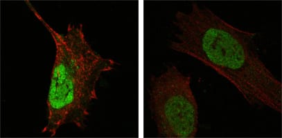
Figure 4: Confocal Immunofluorescence analysis of Hela (left) and L-02 (right) cells using MDM4 mouse mAb (green). Red: Actin filaments have been labeled with DY-554 phalloidin.
For Research Use Only. Not for use in diagnostic procedures.