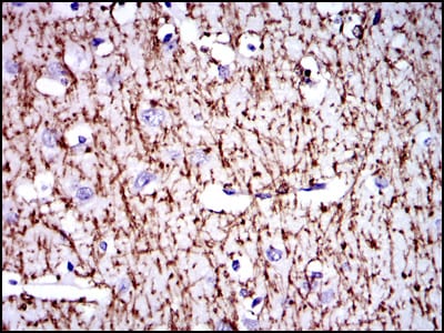
MBP Primary Antibody

Item Information
Catalog #
Size
Price
Description

The protein encoded by the classic MBP gene is a major constituent of the myelin sheath of oligodendrocytes and Schwann cells in the nervous system. However, MBP-related transcripts are also present in the bone marrow and the immune system. These mRNAs arise from the long MBP gene (otherwise called "Golli-MBP") that contains 3 additional exons located upstream of the classic MBP exons. Alternative splicing from the Golli and the MBP transcription start sites gives rise to 2 sets of MBP-related transcripts and gene products. The Golli mRNAs contain 3 exons unique to Golli-MBP, spliced in-frame to 1 or more MBP exons. They encode hybrid proteins that have N-terminal Golli aa sequence linked to MBP aa sequence. The second family of transcripts contain only MBP exons and produce the well characterized myelin basic proteins. This complex gene structure is conserved among species suggesting that the MBP transcription unit is an integral part of the Golli transcription unit and that this arrangement is important for the function and/or regulation of these genes.

Product Overview
Entrez GenelD
4155
Aliases
MGC99675
Clone#
2H9
Host / Isotype
Mouse / IgG1
Species Reactivity
Human
Immunogen
Purified recombinant fragment of human MBP expressed in E. Coli.
Formulation
Ascitic fluid containing 0.03% sodium azide.
Storage
4°C; -20°C for long term storage
Product Applications
WB (Western Blot)
1/500 - 1/2000
IHC_P(Immunohistochemistry)
1/200 - 1/1000
ICC (Immunocytochemistry)
1/200 - 1/1000
FCM (Flow Cytometry)
1/200 - 1/400
ELISA
1/10000
References
1. Cancer Epidemiol Biomarkers Prev. 2009 May;18(5):1651-8.
2. Biochemistry. 2009 Jun 9;48(22):4720-7.
Product Image
Western Blot
Figure 1: Western blot analysis using MBP mAb against human MBP (AA: 1-197) recombinant protein. (Expected MW is 47 kDa)
Western Blot
Figure 2: Western blot analysis using MBP mAb against HEK293 (1) and MBP-hIgGFc transfected HEK293 (2) cell lysate.
Immunohistochemical analysis
Figure 3: Immunohistochemical analysis of paraffin-embedded brain tissues using MBP mouse mAb with DAB staining.
Immunohistochemical analysis
Figure 4: Immunohistochemical analysis of paraffin-embedded cerebellum tissues using MBP mouse mAb with DAB staining.
Immunofluorescence analysis
Figure 5: Immunofluorescence analysis of MSCS cells using MBP mouse mAb (green). Blue: DRAQ5 fluorescent DNA dye. Red: Actin filaments have been labeled with Alexa Fluor-555 phalloidin.
Flow cytometric
Figure 6: Flow cytometric analysis of HepG2 cells using MBP mouse mAb (green) and negative control (red).
Elisa
Black line: Control Antigen (100 ng); Purple line: Antigen(10ng); Blue line: Antigen (50 ng); Red line: Antigen (100 ng);
For Research Use Only. Not for use in diagnostic procedures.