

MARK3 Primary Antibody
Item Information
Catalog #
Size
Price
Description
The protein encoded by this gene is activated by phosphorylation and in turn is involved in the phosphorylation of tau proteins MAP2 and MAP4. Several transcript variants encoding different isoforms have been found for this gene.
Product Overview
Entrez GenelD
4140
Aliases
KP78; CTAK1; PAR1A; Par-1a
Clone#
2G12
Host / Isotype
Mouse / IgG1
Species Reactivity
Human, Mouse
Immunogen
Purified recombinant fragment of human MARK3 (AA: 435-658) expressed in E. Coli.
Formulation
Purified antibody in PBS with 0.05% sodium azide
Storage
Store at 4°C short term. Aliquot and store at -20°C long term. Avoid freeze/thaw cycles.
Product Applications
WB (Western Blot)
1/500 - 1/2000
IHC_P(Immunohistochemistry)
1/200 - 1/1000
FCM (Flow Cytometry)
1/200 - 1/400
ELISA
1/10000
References
1.Biochem Biophys Res Commun. 2010 Apr 16;394(4):890-5. 2.Biochem J. 2008 Apr 15;411(2):249-60.
Product Image
Western Blot
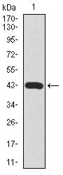
Figure 1: Western blot analysis using MARK3 mAb against human MARK3 recombinant protein. (Expected MW is 40.8 kDa)
Western Blot

Figure 2: Western blot analysis using MARK3 mouse mAb against HeLa (1), SK-N-SH (2), K562 (3), HCT116 (4), HEK293 (5), 3T3L1 (6), NIH3T3 (7), Jurkat (8), and A431 (9) cell lysate.
Flow cytometric

Figure 3: Flow cytometric analysis of SK-N-SH cells using MARK3 mouse mAb (green) and negative control (red).
Immunohistochemical analysis

Figure 4: Immunohistochemical analysis of paraffin-embedded bladder cancer tissues using MARK3 mouse mAb with DAB staining.
Elisa

Black line: Control Antigen (100 ng); Purple line: Antigen(10ng); Blue line: Antigen (50 ng); Red line: Antigen (100 ng);
For Research Use Only. Not for use in diagnostic procedures.