

MAPK3 Primary Antibody
Item Information
Catalog #
Size
Price
Description
The protein encoded by this gene is a member of the MAP kinase family. MAP kinases, also known as extracellular signal-regulated kinases (ERKs), act in a signaling cascade that regulates various cellular processes such as proliferation, differentiation, and cell cycle progression in response to a variety of extracellular signals. This kinase is activated by upstream kinases, resulting in its translocation to the nucleus where it phosphorylates nuclear targets. Alternatively spliced transcript variants encoding different protein isoforms have been described.
Product Overview
Entrez GenelD
5595
Aliases
ERK1; PRKM3; P44ERK1; P44MAPK; HS44KDAP; HUMKER1A; MGC20180
Clone#
1E5
Host / Isotype
Mouse / IgG1
Species Reactivity
Human, Mouse, Rat, Monkey
Immunogen
Purified recombinant fragment of human MAPK3 expressed in E. Coli.
Formulation
Ascitic fluid containing 0.03% sodium azide.
Storage
Store at 4°C short term. Aliquot and store at -20°C long term. Avoid freeze/thaw cycles.
Product Applications
WB (Western Blot)
1/500 - 1/2000
IHC_P(Immunohistochemistry)
1/200 - 1/1000
ICC (Immunocytochemistry)
1/200 - 1/1000
FCM (Flow Cytometry)
1/200 - 1/400
ELISA
1/10000
References
1. Mol Cell. 2009 Nov 13;36(3):477-86.
2. PLoS One. 2009 Oct 22;4(10):e7541.
2. PLoS One. 2009 Oct 22;4(10):e7541.
Product Image
Western Blot

Figure 1: Western blot analysis using MAPK3 mAb against human MAPK3 (AA: 9-143) recombinant protein. (Expected MW is 40.8 kDa)
Western Blot
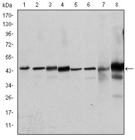
Figure 2: Western blot analysis using MAPK3 mouse mAb against Hela (1), Jurkat (2), RAW264.7 (3), HEK293 (4), K562 (5), NIH/3T3 (6), Cos7 (7) and PC-12 (8) cell lysate.
Immunohistochemical analysis

Figure 3: Immunohistochemical analysis of paraffin-embedded breast cancer tissues using MAPK3 mouse mAb with DAB staining.
Immunohistochemical analysis

Figure 4: Immunohistochemical analysis of paraffin-embedded bladder cancer tissues using MAPK3 mouse mAb with DAB staining.
Immunofluorescence analysis

Figure 5: Immunofluorescence analysis of NIH/3T3 cells using MAPK3 mouse mAb (green). Blue: DRAQ5 fluorescent DNA dye. Red: Actin filaments have been labeled with Alexa Fluor-555 phalloidin.
Flow cytometric

Figure 6: Flow cytometric analysis of Hela cells using MAPK3 mouse mAb (blue) and negative control (red).
Elisa

Red: Control Antigen (100ng); Purple: Antigen (10ng); Green: Antigen (50ng); Blue: Antigen (100ng);
For Research Use Only. Not for use in diagnostic procedures.