

MAPK14 Primary Antibody
Item Information
Catalog #
Size
Price
Description
The protein encoded by this gene is a member of the MAP kinase family. MAP kinases act as an integration point for multiple biochemical signals, and are involved in a wide variety of cellular processes such as proliferation, differentiation, transcription regulation and development. This kinase is activated by various environmental stresses and proinflammatory cytokines. The activation requires its phosphorylation by MAP kinase kinases (MKKs), or its autophosphorylation triggered by the interaction of MAP3K7IP1/TAB1 protein with this kinase. The substrates of this kinase include transcription regulator ATF2, MEF2C, and MAX, cell cycle regulator CDC25B, and tumor suppressor p53, which suggest the roles of this kinase in stress related transcription and cell cycle regulation, as well as in genotoxic stress response. Four alternatively spliced transcript variants of this gene encoding distinct isoforms have been reported.
Product Overview
Entrez GenelD
1432
Aliases
RK; p38; CSBP; EXIP; Mxi2; CSBP1; CSBP2; CSPB1; PRKM14; PRKM15; SAPK2A; p38ALPHA
Clone#
10B11C8
Host / Isotype
Mouse / IgG1
Species Reactivity
Human, Mouse, Monkey, Rat
Immunogen
Purified recombinant fragment of human MAPK14 (AA: 299-360) expressed in E. Coli.
Formulation
Purified antibody in PBS with 0.05% sodium azide
Storage
Store at 4°C short term. Aliquot and store at -20°C long term. Avoid freeze/thaw cycles.
Product Applications
WB (Western Blot)
1/500 - 1/2000
IHC_P(Immunohistochemistry)
1/200 - 1/1000
ELISA
1/10000
References
1. Cytokine. 2012 Oct;60(1):114-21.
2. Clin Cancer Res. 2012 Aug 1;18(15):4037-47.
2. Clin Cancer Res. 2012 Aug 1;18(15):4037-47.
Product Image
Western Blot

Figure 1: Western blot analysis using MAPK14 mAb against human MAPK14 recombinant protein. (Expected MW is 32.6 kDa)
Western Blot
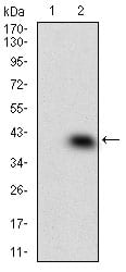
Figure 2: Western blot analysis using MAPK14 mAb against HEK293 (1) and MAPK14 (AA: 299-360)-hIgGFc transfected HEK293 (2) cell lysate.
Western Blot
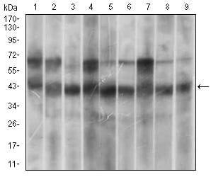
Figure 3: Western blot analysis using MAPK14 mouse mAb against Hela (1), HEK293 (2), A431 (3), MCF-7 (4), RAW264.7 (5), Cos7 (6), C6 (7), Jurkat (8) and NIH/3T3 (9) cell lysate.
Immunohistochemical analysis

Figure 5: Immunohistochemical analysis of paraffin-embedded endometrial cancer tissues using MAPK14 mouse mAb with DAB staining.
Elisa
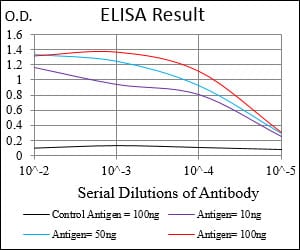
Black line: Control Antigen (100 ng); Purple line: Antigen(10ng); Blue line: Antigen (50 ng); Red line: Antigen (100 ng);
For Research Use Only. Not for use in diagnostic procedures.