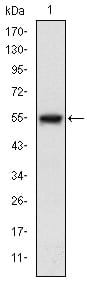
MAP3K5 Primary Antibody

Item Information
Catalog #
Size
Price
Description

Mitogen-activated protein kinase (MAPK) signaling cascades include MAPK or extracellular signal-regulated kinase (ERK), MAPK kinase (MKK or MEK), and MAPK kinase kinase (MAPKKK or MEKK). MAPKK kinase/MEKK phosphorylates and activates its downstream protein kinase, MAPK kinase/MEK, which in turn activates MAPK. The kinases of these signaling cascades are highly conserved, and homologs exist in yeast, Drosophila, and mammalian cells. MAPKKK5 contains 1,374 amino acids with all 11 kinase subdomains. Northern blot analysis shows that MAPKKK5 transcript is abundantly expressed in human heart and pancreas. The MAPKKK5 protein phosphorylates and activates MKK4 (aliases SERK1, MAPKK4) in vitro, and activates c-Jun N-terminal kinase (JNK)/stress-activated protein kinase (SAPK) during transient expression in COS and 293 cells; MAPKKK5 does not activate MAPK/ERK.

Product Overview
Entrez GenelD
4217
Aliases
ASK1; MEKK5; MAPKKK5
Clone#
2E4
Host / Isotype
Mouse / IgG1
Species Reactivity
Human
Immunogen
Purified recombinant fragment of human MAP3K5 expressed in E. Coli.
Formulation
Ascitic fluid containing 0.03% sodium azide.
Storage
4°C; -20°C for long term storage
Product Applications
WB (Western Blot)
1/500 - 1/2000
IHC_P(Immunohistochemistry)
1/200 - 1/1000
ICC (Immunocytochemistry)
1/200 - 1/1000
FCM (Flow Cytometry)
1/200 - 1/400
ELISA
1/10000
References
1. Mol Cancer. 2009 Dec 27;8:130.
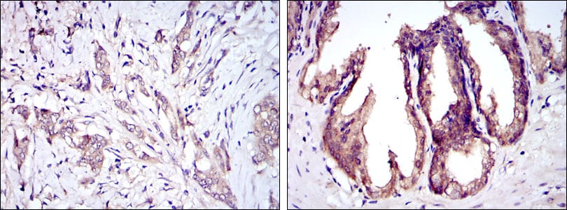
2. J Biol Chem. 2009 Oct 30;284(44):30372-82.
Product Image
Western Blot
Figure 1: Western blot analysis using MAP3K5 mAb against human MAP3K5 (AA: 922-1108) recombinant protein. (Expected MW is 46.5 kDa)
Immunohistochemical analysis
Figure 2: Immunohistochemical analysis of paraffin-embedded breast cancer tissues (left) and prostate tissues (right) using MAP3K5 mouse mAb with DAB staining.
Immunofluorescence analysis
Figure 3: Immunofluorescence analysis of Hela cells using MAP3K5 mouse mAb (green). Blue: DRAQ5 fluorescent DNA dye. Red: Actin filaments have been labeled with Alexa Fluor-555 phalloidin.
Flow cytometric
Figure 4: Flow cytometric analysis of Hela cells using MAP3K5 mouse mAb (green) and negative control (purple).
Elisa
Red: Control Antigen (100ng); Purple: Antigen (10ng); Green: Antigen (50ng); Blue: Antigen (100ng);
For Research Use Only. Not for use in diagnostic procedures.