
MAP2K5 Primary Antibody
Item Information
Catalog #
Size
Price
Description
The protein encoded by this gene is a dual specificity protein kinase that belongs to the MAP kinase kinase family. This kinase specifically interacts with and activates MAPK7/ERK5. This kinase itself can be phosphorylated and activated by MAP3K3/MEKK3, as well as by atypical protein kinase C isoforms (aPKCs). The signal cascade mediated by this kinase is involved in growth factor stimulated cell proliferation and muscle cell differentiation. Three alternatively spliced transcript variants of this gene encoding distinct isoforms have been described.
Product Overview
Entrez GenelD
5607
Aliases
MEK5; MAPKK5; PRKMK5; HsT17454
Clone#
5H11B10
Host / Isotype
Mouse / IgG1
Species Reactivity
Human
Immunogen
Purified recombinant fragment of human MAP2K5 (AA: 63-180) expressed in E. Coli.
Formulation
Purified antibody in PBS with 0.05% sodium azide.
Storage
Store at 4°C short term. Aliquot and store at -20°C long term. Avoid freeze/thaw cycles.
Product Applications
WB (Western Blot)
1/500 - 1/2000
IHC_P(Immunohistochemistry)
1/200 - 1/1000
FCM (Flow Cytometry)
1/200 - 1/400
ELISA
1/10000
References
1. DNA Cell Biol. 2012 Mar;31(3):342-9.
2. Wei Sheng Yan Jiu. 2006 Mar;35(2):184-6.
2. Wei Sheng Yan Jiu. 2006 Mar;35(2):184-6.
Product Image
Western Blot

Figure 1: Western blot analysis using MAP2K5 mAb against human MAP2K5 (AA: 63-180) recombinant protein. (Expected MW is 39 kDa)
Western Blot
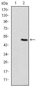
Figure 2: Western blot analysis using MAP2K5 mAb against HEK293 (1) and MAP2K5 (AA: 63-180)-hIgGFc transfected HEK293 (2) cell lysate.
Western Blot

Figure 3: Western blot analysis using MAP2K5 mouse mAb against Jurkat (1), A431 (2), A549 (3) cell lysate.
Flow cytometric

Figure 4: Flow cytometric analysis of Jurkat cells using MAP2K5 mouse mAb (green) and negative control (red).
Immunohistochemical analysis

Figure 5: Immunohistochemical analysis of paraffin-embedded liver cancer tissues using MAP2K5 mouse mAb with DAB staining.
Elisa

Black line: Control Antigen (100 ng); Purple line: Antigen(10ng); Blue line: Antigen (50 ng); Red line: Antigen (100 ng);
For Research Use Only. Not for use in diagnostic procedures.