
MAP2K3 Primary Antibody
Item Information
Catalog #
Size
Price
Description
The protein encoded by this gene is a dual specificity protein kinase that belongs to the MAP kinase kinase family. This kinase is activated by mitogenic and environmental stress, and participates in the MAP kinase-mediated signaling cascade. It phosphorylates and thus activates MAPK14/p38-MAPK. This kinase can be activated by insulin, and is necessary for the expression of glucose transporter. Expression of RAS oncogene is found to result in the accumulation of the active form of this kinase, which thus leads to the constitutive activation of MAPK14, and confers oncogenic transformation of primary cells. The inhibition of this kinase is involved in the pathogenesis of Yersina pseudotuberculosis. Multiple alternatively spliced transcript variants that encode distinct isoforms have been reported for this gene.
Product Overview
Entrez GenelD
5606
Aliases
MEK3; MKK3; MAPKK3; PRKMK3; SAPKK2; SAPKK-2
Clone#
2E12D11
Host / Isotype
Mouse / IgG1
Species Reactivity
Human
Immunogen
Purified recombinant fragment of human MAP2K3 (AA: 1-138) expressed in E. Coli.
Formulation
Purified antibody in PBS with 0.05% sodium azide
Storage
Store at 4°C short term. Aliquot and store at -20°C long term. Avoid freeze/thaw cycles.
Product Applications
WB (Western Blot)
1/500 - 1/2000
IHC_P(Immunohistochemistry)
1/200 - 1/1000
ICC (Immunocytochemistry)
1/200 - 1/1000
FCM (Flow Cytometry)
1/200 - 1/400
ELISA
1/10000
References
1.Hum Mol Genet. 2013 Nov 1;22(21):4438-49.
2.Proteomics Clin Appl. 2010 Nov;4(10-11):816-28.
2.Proteomics Clin Appl. 2010 Nov;4(10-11):816-28.
Product Image
Elisa
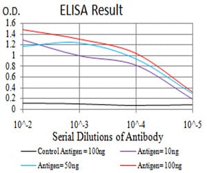
Figure 1: Black line: Control Antigen (100 ng);Purple line: Antigen (10ng); Blue line: Antigen (50 ng); Red line:Antigen (100 ng)
Western Blot
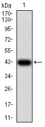
Figure 2:Western blot analysis using MAP2K3 mAb against human MAP2K3 (AA: 1-138) recombinant protein. (Expected MW is 42.1 kDa)
Western Blot
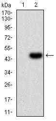
Figure 3:Western blot analysis using MAP2K3 mAb against HEK293 (1) and MAP2K3 (AA: 1-138)-hIgGFc transfected HEK293 (2) cell lysate.
Immunofluorescence analysis
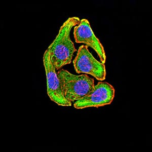
Figure 4:Immunofluorescence analysis of GC-7901 cells using MAP2K3 mouse mAb (green). Blue: DRAQ5 fluorescent DNA dye. Red: Actin filaments have been labeled with Alexa Fluor- 555 phalloidin. Secondary antibody from Fisher (Cat#: 35503)
Immunofluorescence analysis
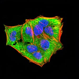
Figure 5:Immunofluorescence analysis of Hela cells using MAP2K3 mouse mAb (green). Blue: DRAQ5 fluorescent DNA dye. Red: Actin filaments have been labeled with Alexa Fluor- 555 phalloidin. Secondary antibody from Fisher (Cat#: 35503)
Immunofluorescence analysis
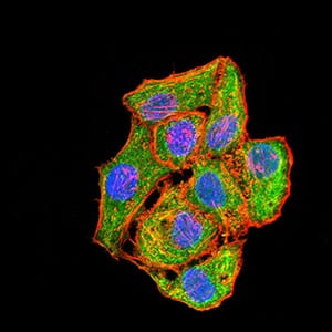
Figure 6:Immunofluorescence analysis of HepG2 cells using MAP2K3 mouse mAb (green). Blue: DRAQ5 fluorescent DNA dye. Red: Actin filaments have been labeled with Alexa Fluor- 555 phalloidin. Secondary antibody from Fisher (Cat#: 35503)
Flow cytometric
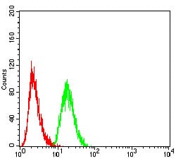
Figure 7:Flow cytometric analysis of Hela cells using MAP2K3 mouse mAb (green) and negative control (red).
Immunohistochemical analysis
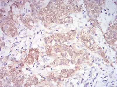
Figure 8:Immunohistochemical analysis of paraffin-embedded bladder cancer tissues using MAP2K3 mouse mAb with DAB staining.
Immunohistochemical analysis
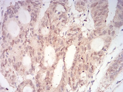
Figure 9:Immunohistochemical analysis of paraffin-embedded rectum cancer tissues using MAP2K3 mouse mAb with DAB staining.
For Research Use Only. Not for use in diagnostic procedures.