

MAP1LC3A Primary Antibody
Item Information
Catalog #
Size
Price
Description
MAP1A and MAP1B are microtubule-associated proteins which mediate the physical interactions between microtubules and components of the cytoskeleton. MAP1A and MAP1B each consist of a heavy chain subunit and multiple light chain subunits. The protein encoded by this gene is one of the light chain subunits and can associate with either MAP1A or MAP1B. Two transcript variants encoding different isoforms have been found for this gene. The expression of variant 1 is suppressed in many tumor cell lines, suggesting that may be involved in carcinogenesis.
Product Overview
Entrez GenelD
84557
Aliases
LC3; LC3A; ATG8E; MAP1ALC3; MAP1BLC3
Clone#
5B10D8
Host / Isotype
Mouse / IgG1
Species Reactivity
Human
Immunogen
Purified recombinant fragment of human MAP1LC3A (AA: 1-121) expressed in E. Coli.
Formulation
Purified antibody in PBS with 0.05% sodium azide
Storage
Store at 4°C short term. Aliquot and store at -20°C long term. Avoid freeze/thaw cycles.
Product Applications
WB (Western Blot)
1/500 - 1/2000
IHC_P(Immunohistochemistry)
1/200 - 1/1000
FCM (Flow Cytometry)
1/200 - 1/400
ELISA
1/10000
References
1.Mol Biol Rep. 2012 Jan;39(1):259-67.
2.Tohoku J Exp Med. 2011;223(4):243-51.
2.Tohoku J Exp Med. 2011;223(4):243-51.
Product Image
Western Blot

Figure 1: Western blot analysis using MAP1LC3A mAb against human MAP1LC3A recombinant protein. (Expected MW is 39.8 kDa)
Western Blot

Figure 2: Western blot analysis using MAP1LC3A mAb against HEK293 (1) and MAP1LC3A (AA: 1-121)-hIgGFc transfected HEK293 (2) cell lysate.
Flow cytometric
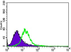
Figure 3: Flow cytometric analysis of HeLa cells using MAP1LC3A mouse mAb (green) and negative control (purple).
Immunohistochemical analysis

Figure 4: Immunohistochemical analysis of paraffin-embedded liver cancer tissues using MAP1LC3A mouse mAb with DAB staining.
Immunohistochemical analysis

Figure 5: Immunohistochemical analysis of paraffin-embedded testis tissues using MAP1LC3A mouse mAb with DAB staining.
Elisa

Black line: Control Antigen (100 ng); Purple line: Antigen(10ng); Blue line: Antigen (50 ng); Red line: Antigen (100 ng);
For Research Use Only. Not for use in diagnostic procedures.