

LRP3 Primary Antibody
Item Information
Catalog #
Size
Price
Description
LRP3 (LDL Receptor Related Protein 3) is a Protein Coding genewhich may be involved in the internalization of lipophilic molecules and/or signal transduction. Its precise role is however unclear, since it does not bind to very low density lipoprotein (VLDL) or to LRPAP1 in vitro.
Product Overview
Entrez GenelD
4037
Clone#
7B3A5E9
Host / Isotype
Mouse / IgG1
Species Reactivity
Human
Immunogen
Purified recombinant fragment of human LRP3 (AA: extra 43-184) expressed in E. Coli.
Formulation
Purified antibody in PBS with 0.05% sodium azide
Storage
Store at 4°C short term. Aliquot and store at -20°C long term. Avoid freeze/thaw cycles.
Product Applications
WB (Western Blot)
1/500 - 1/2000
ICC (Immunocytochemistry)
1/100 - 1/500
FCM (Flow Cytometry)
1/200 - 1/400
ELISA
1/10000
References
1.Stem Cell Res. 2017 Apr;20:94-104.
Product Image
Elisa
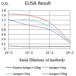
Figure 1:Black line: Control Antigen (100 ng);Purple line: Antigen (10ng); Blue line: Antigen (50 ng); Red line:Antigen (100 ng)
Western Blot
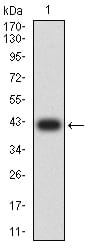
Figure 2:Western blot analysis using LRP3 mAb against human LRP3 (AA: extra 43-184) recombinant protein. (Expected MW is 41.7 kDa)
Western Blot
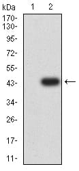
Figure 3:Western blot analysis using LRP3 mAb against HEK293 (1) and LRP3 (AA: extra 43-184)-hIgGFc transfected HEK293 (2) cell lysate.
Western Blot
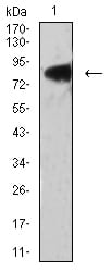
Figure 4:Western blot analysis using LRP3 mouse mAb against PANC-1 (1) cell lysate.
Immunofluorescence analysis
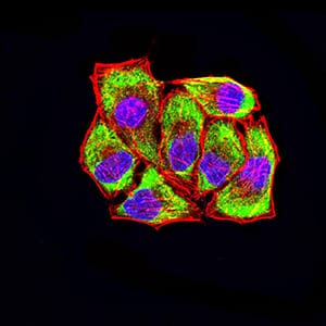
Figure 5:Immunofluorescence analysis of Hela cells using LRP3 mouse mAb (green). Blue: DRAQ5 fluorescent DNA dye. Red: Actin filaments have been labeled with Alexa Fluor- 555 phalloidin. Secondary antibody from Fisher (Cat#: 35503)
Flow cytometric
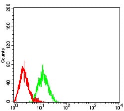
Figure 6:Flow cytometric analysis of HL-60 cells using LRP3 mouse mAb (green) and negative control (red).
For Research Use Only. Not for use in diagnostic procedures.