
LRP1 Primary Antibody
Item Information
Catalog #
Size
Price
Description
The protein encoded by this gene is an endocytic receptor involved in several cellular processes, including intracellular signaling, lipid homeostasis, and clearance of apoptotic cells. In addition, the encoded protein is necessary for the A2M-mediated clearance of secreted amyloid precursor protein and beta-amyloid, the main component of amyloid plaques found in Alzheimer patients. Expression of this gene decreases with age and has been found to be lower than controls in brain tissue from Alzheimer patients.
Product Overview
Entrez GenelD
4035
Aliases
APR; LRP; A2MR; CD91; APOER; LRP1A; TGFBR5; IGFBP3R
Clone#
1F6C6
Host / Isotype
Mouse / IgG1
Species Reactivity
Human
Immunogen
Purified recombinant fragment of human LRP1 (AA: 20-155) expressed in E. Coli.
Formulation
Purified antibody in PBS with 0.05% sodium azide
Storage
Store at 4°C short term. Aliquot and store at -20°C long term. Avoid freeze/thaw cycles.
Product Applications
WB (Western Blot)
1/500 - 1/2000
ICC (Immunocytochemistry)
1/200 - 1/1000
FCM (Flow Cytometry)
1/200 - 1/400
ELISA
1/10000
References
1.Biomed Res Int. 2013;2013:152163.
2.J Transl Med. 2012 Aug 8;10:160.
2.J Transl Med. 2012 Aug 8;10:160.
Product Image
Elisa

Figure 1: Black line: Control Antigen (100 ng); Purple line: Antigen(10ng); Blue line: Antigen (50 ng); Red line: Antigen (100 ng);
Western Blot
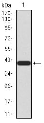
Figure 2:Western blot analysis using LRP1 mAb against human LRP1 (AA: 20-155) recombinant protein. (Expected MW is 40.8 kDa)
Western Blot

Figure 3:Western blot analysis using LRP1 mAb against HEK293 (1) and LRP1 (AA: 20-155)-hIgGFc transfected HEK293 (2) cell lysate.
Immunofluorescence analysis
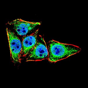
Figure 4:Immunofluorescence analysis of HeLa cells using LRP1 mouse mAb (green). Blue: DRAQ5 fluorescent DNA dye. Red: Actin filaments have been labeled with Alexa Fluor- 555 phalloidin. Secondary antibody from Fisher (Cat#: 35503)
Flow cytometric

Figure 5:Flow cytometric analysis of Raji cells using LRP1 mouse mAb (green) and negative control (red).
For Research Use Only. Not for use in diagnostic procedures.