

LPlunc1 Primary Antibody
Item Information
Catalog #
Size
Price
Description
The protein encoded by this gene may be involved in the innate immune response to bacterial exposure in the mouth, nasal cavities, and lungs. The encoded protein is secreted and is a member of the BPI/LBP/PLUNC protein superfamily. This gene is found with other members of the superfamily in a cluster on chromosome 20.Â
Product Overview
Entrez GenelD
92747
Aliases
BPIFB1; LPLUNC1; MGC14597; C20orf114
Clone#
2A5
Host / Isotype
Mouse / IgG1
Species Reactivity
Human
Immunogen
Purified recombinant fragment of human LPlunc1 expressed in E. Coli.
Formulation
Purified antibody in PBS with 0.05% sodium azide
Storage
Store at 4°C short term. Aliquot and store at -20°C long term. Avoid freeze/thaw cycles.
Product Applications
WB (Western Blot)
1/500 - 1/2000
IHC_P(Immunohistochemistry)
1/200 - 1/1000
ICC (Immunocytochemistry)
1/200 - 1/1000
FCM (Flow Cytometry)
1/200 - 1/400
ELISA
1/10000
References
1. Genes Immun. 2009 Apr;10(3):267-72.
2. Histochem Cell Biol. 2010 May;133(5):505-15.
2. Histochem Cell Biol. 2010 May;133(5):505-15.
Product Image
Western Blot

Figure 1: Western blot analysis using LPlunc1 mAb against human LPlunc1 recombinant protein. (Expected MW is 52 kDa)
Immunohistochemical analysis

Figure 2: Immunohistochemical analysis of paraffin-embedded brain tissues using LPlunc1 mouse mAb with DAB staining.
Immunohistochemical analysis

Figure 3: Immunohistochemical analysis of paraffin-embedded tonsil tissues using LPlunc1 mouse mAb with DAB staining.
Immunofluorescence analysis

Figure 4: Immunofluorescence analysis of Hela cells using LPlunc1 mouse mAb (green). Blue: DRAQ5 fluorescent DNA dye. Red: Actin filaments have been labeled with Alexa Fluor-555 phalloidin.
Flow cytometric
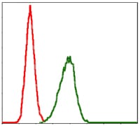
Figure 5: Flow cytometric analysis of Hela cells using LPlunc1 mouse mAb (green) and negative control (red).
Elisa

Black line: Control Antigen (100 ng); Purple line: Antigen(10ng); Blue line: Antigen (50 ng); Red line: Antigen (100 ng);
For Research Use Only. Not for use in diagnostic procedures.