

LPA Primary Antibody
Item Information
Catalog #
Size
Price
Description
The protein encoded by this gene is a serine proteinase that inhibits the activity of tissue-type plasminogen activator I. The encoded protein constitutes a substantial portion of lipoprotein(a) and is proteolytically cleaved, resulting in fragments that attach to atherosclerotic lesions and promote thrombogenesis. Elevated plasma levels of this protein are linked to atherosclerosis. Depending on the individual, the encoded protein contains 2-43 copies of kringle-type domains. The allele represented here contains 15 copies of the kringle-type repeats and corresponds to that found in the reference genome sequence.
Product Overview
Entrez GenelD
4018
Aliases
LP; AK38; APOA
Clone#
4H1
Host / Isotype
Mouse / IgG1
Species Reactivity
Human
Immunogen
Purified recombinant fragment of human LPA (AA:1823-2013) expressed in E. Coli.
Formulation
Purified antibody in PBS with 0.05% sodium azide
Storage
Store at 4°C short term. Aliquot and store at -20°C long term. Avoid freeze/thaw cycles.
Product Applications
WB (Western Blot)
1/500 - 1/2000
IHC_P(Immunohistochemistry)
1/200 - 1/1000
ICC (Immunocytochemistry)
1/200 - 1/1000
ELISA
1/10000
References
1.J Lipid Res. 2010 Oct;51(10):3055-61.
2.Thromb Res. 2010 Sep;126(3):222-6.
2.Thromb Res. 2010 Sep;126(3):222-6.
Product Image
Western Blot

Figure 1: Western blot analysis using LPA mAb against human LPA recombinant protein. (Expected MW is 34.1 kDa)
Immunohistochemical analysis
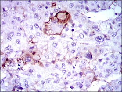
Figure 2: Immunohistochemical analysis of paraffin-embedded liver cancer tissues using LPA mouse mAb with DAB staining.
Immunohistochemical analysis

Figure 3: Immunohistochemical analysis of paraffin-embedded breast cancer tissues using LPA mouse mAb with DAB staining.
Immunohistochemical analysis

Figure 4: Immunohistochemical analysis of paraffin-embedded rectum cancer tissues using LPA mouse mAb with DAB staining.
Immunofluorescence analysis

Figure 5: Immunofluorescence analysis of HepG2 cells using LPA mouse mAb (green). Blue: DRAQ5 fluorescent DNA dye. Red: Actin filaments have been labeled with Alexa Fluor-555 phalloidin.
Elisa

Black line: Control Antigen (100 ng); Purple line: Antigen(10ng); Blue line: Antigen (50 ng); Red line: Antigen (100 ng);
For Research Use Only. Not for use in diagnostic procedures.