
LMNB2 Primary Antibody
Item Information
Catalog #
Size
Price
Description
This gene encodes a B type nuclear lamin. The nuclear lamina consists of a two-dimensional matrix of proteins located next to the inner nuclear membrane. The lamin family of proteins make up the matrix and are highly conserved in evolution. During mitosis, the lamina matrix is reversibly disassembled as the lamin proteins are phosphorylated. Lamin proteins are thought to be involved in nuclear stability, chromatin structure and gene expression. Vertebrate lamins consist of two types, A and B. Mutations in this gene are associated with acquired partial lipodystrophy.
Product Overview
Entrez GenelD
84823
Aliases
LMN2; LAMB2
Clone#
2E2F4
Host / Isotype
Mouse / IgG2b
Species Reactivity
Human
Immunogen
Purified recombinant fragment of human LMNB2 (AA: 401-600) expressed in E. Coli.
Formulation
Purified antibody in PBS with 0.05% sodium azide
Storage
Store at 4°C short term. Aliquot and store at -20°C long term. Avoid freeze/thaw cycles.
Product Applications
WB (Western Blot)
1/500 - 1/2000
IHC_P(Immunohistochemistry)
1/200 - 1/1000
ICC (Immunocytochemistry)
1/200 - 1/1000
FCM (Flow Cytometry)
1/200 - 1/400
ELISA
1/10000
References
1.J Pediatr Endocrinol Metab. 2012;25(3-4):375-7.
2.FEBS Lett. 2006 Nov 13;580(26):6211-6.
2.FEBS Lett. 2006 Nov 13;580(26):6211-6.
Product Image
Western Blot

Figure 2:Western blot analysis using LMNB2 mAb against human LMNB2 (AA: 401-600) recombinant protein. (Expected MW is 47.6 kDa)
Western Blot

Figure 3:Western blot analysis using LMNB2 mAb against HEK293 (1) and LMNB2 (AA: 401-600)-hIgGFc transfected HEK293 (2) cell lysate.
Western Blot
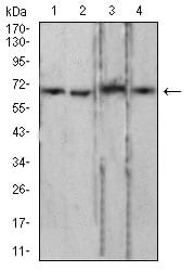
Figure 4:Western blot analysis using LMNB2 mouse mAb against PC-3 (1), LNcap (2), Jurkat (3), and HCT116 (4) cell lysate.
Immunofluorescence analysis

Figure 5:Immunofluorescence analysis of GC-7901 cells using LMNB2 mouse mAb (green). Blue: DRAQ5 fluorescent DNA dye. Red: Actin filaments have been labeled with Alexa Fluor- 555 phalloidin. Secondary antibody from Fisher (Cat#: 35503)
Immunofluorescence analysis

Figure 6:Immunofluorescence analysis of Hela cells using LMNB2 mouse mAb (green). Blue: DRAQ5 fluorescent DNA dye. Red: Actin filaments have been labeled with Alexa Fluor- 555 phalloidin. Secondary antibody from Fisher (Cat#: 35503)
Immunofluorescence analysis

Figure 7:Immunofluorescence analysis of HepG2 cells using LMNB2 mouse mAb (green). Blue: DRAQ5 fluorescent DNA dye. Red: Actin filaments have been labeled with Alexa Fluor- 555 phalloidin. Secondary antibody from Fisher (Cat#: 35503)
Flow cytometric

Figure 8:Flow cytometric analysis of Hela cells using LMNB2 mouse mAb (green) and negative control (red).
Immunohistochemical analysis

Figure 9:Immunohistochemical analysis of paraffin-embedded colon cancer tissues using LMNB2 mouse mAb with DAB staining.
Immunohistochemical analysis

Figure 10:Immunohistochemical analysis of paraffin-embedded ovarian cancer tissues using LMNB2 mouse mAb with DAB staining.
Elisa

Black line: Control Antigen (100 ng);Purple line: Antigen (10ng); Blue line: Antigen (50 ng); Red line:Antigen (100 ng)
For Research Use Only. Not for use in diagnostic procedures.