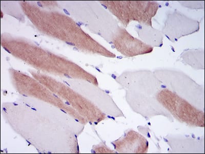
LMNA Primary Antibody

Item Information
Catalog #
Size
Price
Description

The nuclear lamina consists of a two-dimensional matrix of proteins located next to the inner nuclear membrane. The lamin family of proteins make up the matrix and are highly conserved in evolution. During mitosis, the lamina matrix is reversibly disassembled as the lamin proteins are phosphorylated. Lamin proteins are thought to be involved in nuclear stability, chromatin structure and gene expression. Vertebrate lamins consist of two types, A and B. Through alternate splicing, this gene encodes three type A lamin isoforms. Mutations in this gene lead to several diseases: Emery-Dreifuss muscular dystrophy, familial partial lipodystrophy, limb girdle muscular dystrophy, dilated cardiomyopathy, Charcot-Marie-Tooth disease, and Hutchinson-Gilford progeria syndrome.

Product Overview
Entrez GenelD
4000
Aliases
FPL; IDC; LFP; CDDC; EMD2; FPLD; HGPS; LDP1; LMN1; LMNC; PRO1; CDCD1; CMD1A; FPLD2; LMNL1; CMT2B1
Clone#
4E7
Host / Isotype
Mouse / IgG1
Species Reactivity
Human, Mouse, Rat
Immunogen
Purified recombinant fragment of human LMNA expressed in E. Coli.
Formulation
Ascitic fluid containing 0.03% sodium azide.
Storage
4°C; -20°C for long term storage
Product Applications
WB (Western Blot)
1/500 - 1/2000
IHC_P(Immunohistochemistry)
1/200 - 1/1000
ELISA
1/10000
References
1. Muscle Nerve. 2010 Jan;41(1):85-91.
2. Endocrine. 2009 Dec;36(3):518-23.
Product Image
Western Blot
Figure 1: Western blot analysis using LMNA mAb against human LMNA (AA: 212-477) recombinant protein. (Expected MW is 56.3 kDa)
Western Blot
Figure 2: Western blot analysis using LMNA mouse mAb against Raw264.7 (1), PC-12 (2), THP-1 (3), A431 (4), MCF-7 (5) and Jurkat (6) cell lysate.
Immunohistochemical analysis
Figure 3: Immunohistochemical analysis of paraffin-embedded human striated muscle tissues using LMNA mouse mAb with DAB staining.
Elisa
Red: Control Antigen (100ng); Purple: Antigen (10ng); Green: Antigen (50ng); Blue: Antigen (100ng);
For Research Use Only. Not for use in diagnostic procedures.