

LILRA3 Primary Antibody
Item Information
Catalog #
Size
Price
Description
This gene encodes a member of a family of immunoreceptors that are expressed predominantly in monocytes and B cells, and at lower levels in dendritic cells and natural killer cells. The encoded protein lacks the transmembrane region found in other members of this family. It acts as a soluble receptor for class I major histocompatibility complex (MHC) antigens. Alternatively spliced transcript variants encoding different isoforms have been found. This gene is located in a cluster of related genes on chromosome 19 and is polymorphic in human populations, with many individuals containing a deletion of this genomic region.
Product Overview
Entrez GenelD
11026
Aliases
HM31; HM43; ILT6; LIR4; CD85E; ILT-6; LIR-4
Clone#
6F12D7
Host / Isotype
Mouse / IgG1
Species Reactivity
Human
Immunogen
Purified recombinant fragment of human LILRA3 (AA: 24-168) expressed in E. Coli.
Formulation
Purified antibody in PBS with 0.05% sodium azide
Storage
Store at 4°C short term. Aliquot and store at -20°C long term. Avoid freeze/thaw cycles.
Product Applications
WB (Western Blot)
1/500 - 1/2000
FCM (Flow Cytometry)
1/200 - 1/400
ELISA
1/10000
References
1.Clin Exp Rheumatol. 2016 Sep-Oct;34 Suppl 100(5):208-209. 2.PLoS One. 2016 Feb 12;11(2):e0149200.
Product Image
Elisa
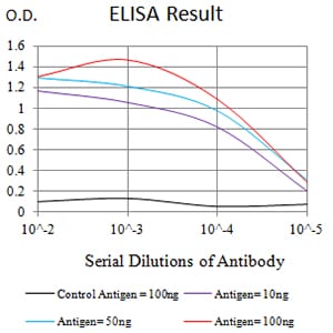
Figure 1: Black line: Control Antigen (100 ng);Purple line: Antigen (10ng); Blue line: Antigen (50 ng); Red line:Antigen (100 ng)
Western Blot

Figure 2:Western blot analysis using LILRA3 mAb against human LILRA3 (AA: 24-168) recombinant protein. (Expected MW is 41.7 kDa)
Western Blot

Figure 3:Western blot analysis using LILRA3 mAb against HEK293 (1) and LILRA3 (AA: 24-168)-hIgGFc transfected HEK293 (2) cell lysate.
Flow cytometric
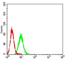
Figure 4:Flow cytometric analysis of K562 cells using LILRA3 mouse mAb (green) and negative control (red).
For Research Use Only. Not for use in diagnostic procedures.