
LILRA2 Primary Antibody
Item Information
Catalog #
Size
Price
Description
This gene encodes a member of a family of immunoreceptors that are expressed predominantly on monocytes and B cells, and at lower levels on dendritic cells and natural killer cells. The encoded protein is an activating receptor that inhibits dendritic cell differentiation and antigen presentation and suppresses innate immune response. Alternatively spliced transcript variants encoding different isoforms have been found. This gene is located in a cluster of related genes on chromosome 19 and there is a pseudogene for this gene on chromosome 3.
Product Overview
Entrez GenelD
11027
Aliases
ILT1; LIR7; CD85H; LIR-7
Clone#
2G5E12
Host / Isotype
Mouse / IgG1
Species Reactivity
Human
Immunogen
Purified recombinant fragment of human LILRA2 (AA: extra 316-449) expressed in E. Coli.
Formulation
Purified antibody in PBS with 0.05% sodium azide
Storage
Store at 4°C short term. Aliquot and store at -20°C long term. Avoid freeze/thaw cycles.
Product Applications
WB (Western Blot)
1/500 - 1/2000
FCM (Flow Cytometry)
1/200 - 1/400
ELISA
1/10000
References
1.Nat Microbiol. 2016 Apr 25;1(6):16054. 2.PLoS One. 2012;7(3):e33478.
Product Image
Elisa
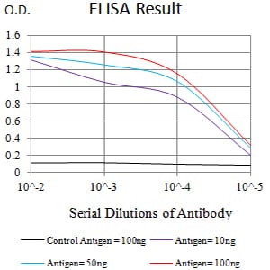
Figure 1:Black line: Control Antigen (100 ng);Purple line: Antigen (10ng); Blue line: Antigen (50 ng); Red line:Antigen (100 ng)
Western Blot
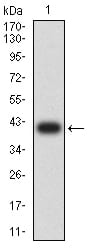
Figure 2:Western blot analysis using LILRA2 mAb against human LILRA2 (AA: extra 316-449) recombinant protein. (Expected MW is 40.6 kDa)
Western Blot
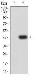
Figure 3:Western blot analysis using LILRA2 mAb against HEK293 (1) and LILRA2 (AA: extra 316-449)-hIgGFc transfected HEK293 (2) cell lysate.
Flow cytometric
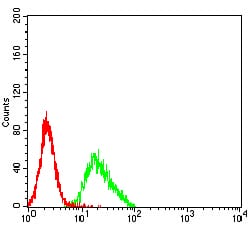
Figure 4:Flow cytometric analysis of K562 cells using LILRA2 mouse mAb (green) and negative control (red).
For Research Use Only. Not for use in diagnostic procedures.