
LEF1 Primary Antibody
Item Information
Catalog #
Size
Price
Description
This gene encodes a transcription factor belonging to a family of proteins that share homology with the high mobility group protein-1. The protein encoded by this gene can bind to a functionally important site in the T-cell receptor-alpha enhancer, thereby conferring maximal enhancer activity. This transcription factor is involved in the Wnt signaling pathway, and it may function in hair cell differentiation and follicle morphogenesis. Mutations in this gene have been found in somatic sebaceous tumors. This gene has also been linked to other cancers, including androgen-independent prostate cancer. Alternative splicing results in multiple transcript variants.
Product Overview
Entrez GenelD
51176
Aliases
LEF-1; TCF10; TCF7L3; TCF1ALPHA
Clone#
1A4B11
Host / Isotype
Mouse / IgG1
Species Reactivity
Human
Immunogen
Purified recombinant fragment of human LEF1 (AA: 33-138) expressed in E. Coli.
Formulation
Purified antibody in PBS with 0.05% sodium azide
Storage
Store at 4°C short term. Aliquot and store at -20°C long term. Avoid freeze/thaw cycles.
Product Applications
WB (Western Blot)
1/500 - 1/2000
IHC_P(Immunohistochemistry)
1/200 - 1/1000
ELISA
1/10000
References
1.Cancer Invest. 2014 Aug;32(7):368-74.
2.BMC Gastroenterol. 2012 May 28;12:53.
2.BMC Gastroenterol. 2012 May 28;12:53.
Product Image
Elisa
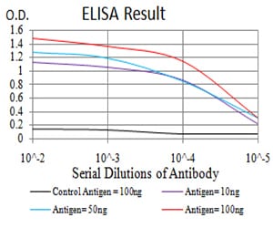
Figure 1: Black line: Control Antigen (100 ng); Purple line: Antigen(10ng); Blue line: Antigen (50 ng); Red line: Antigen (100 ng);
Western Blot
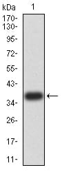
Figure 2:Western blot analysis using LEF1 mAb against human LEF1 (AA: 33-138) recombinant protein. (Expected MW is 37.1 kDa)
Western Blot
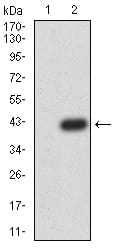
Figure 3:Western blot analysis using LEF1 mAb against HEK293 (1) and LEF1 (AA: 33-138)-hIgGFc transfected HEK293 (2) cell lysate.
Western Blot
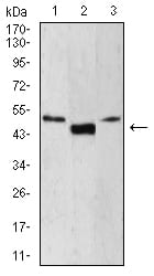
Figure 4:Western blot analysis using LEF1 mouse mAb against Jurkat (1), HepG2 (2), and MOLT4 (3) cell lysate.
Immunohistochemical analysis
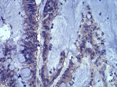
Figure 5:Immunohistochemical analysis of paraffin-embedded colon cancer tissues using LEF1 mouse mAb with DAB staining.
Immunohistochemical analysis
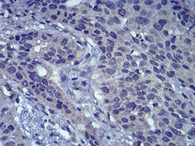
Figure 6:Immunohistochemical analysis of paraffin-embedded esophageal cancer tissues using LEF1 mouse mAb with DAB staining.
For Research Use Only. Not for use in diagnostic procedures.