
LDLR Primary Antibody
Item Information
Catalog #
Size
Price
Description
The low density lipoprotein receptor (LDLR) gene family consists of cell surface proteins involved in receptor-mediated endocytosis of specific ligands. Low density lipoprotein (LDL) is normally bound at the cell membrane and taken into the cell ending up in lysosomes where the protein is degraded and the cholesterol is made available for repression of microsomal enzyme 3-hydroxy-3-methylglutaryl coenzyme A (HMG CoA) reductase, the rate-limiting step in cholesterol synthesis. At the same time, a reciprocal stimulation of cholesterol ester synthesis takes place. Mutations in this gene cause the autosomal dominant disorder, familial hypercholesterolemia. Alternate splicing results in multiple transcript variants.r
Product Overview
Entrez GenelD
3949
Aliases
FH; FHC; LDLCQ2
Clone#
1B10H10
Host / Isotype
Mouse / IgG1
Species Reactivity
Human
Immunogen
Purified recombinant fragment of human LDLR (AA: 22-150) expressed in E. Coli.
Formulation
Purified antibody in PBS with 0.05% sodium azide
Storage
Store at 4°C short term. Aliquot and store at -20°C long term. Avoid freeze/thaw cycles.
Product Applications
WB (Western Blot)
1/500 - 1/2000
IHC_P(Immunohistochemistry)
1/200 - 1/1000
FCM (Flow Cytometry)
1/200 - 1/400
ELISA
1/10000
References
Phytother Res. 2012 Nov;26(11):1688-94.
Biochim Biophys Acta. 2011 Jun;1811(6):397-408.
Biochim Biophys Acta. 2011 Jun;1811(6):397-408.
Product Image
Elisa

Figure 1: Black line: Control Antigen (100 ng); Purple line: Antigen(10ng); Blue line: Antigen (50 ng); Red line: Antigen (100 ng);
Western Blot

Figure 2:Western blot analysis using LDLR mAb against human LDLR (AA: 22-150) recombinant protein. (Expected MW is 40.1 kDa)
Western Blot

Figure 3:Western blot analysis using LDLR mAb against HEK293 (1) and LDLR (AA: 22-150)-hIgGFc transfected HEK293 (2) cell lysate.
Flow cytometric
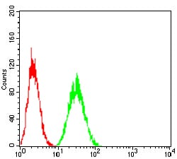
Figure 4:Flow cytometric analysis of Hela cells using LDLR mouse mAb (green) and negative control (red).
Immunohistochemical analysis
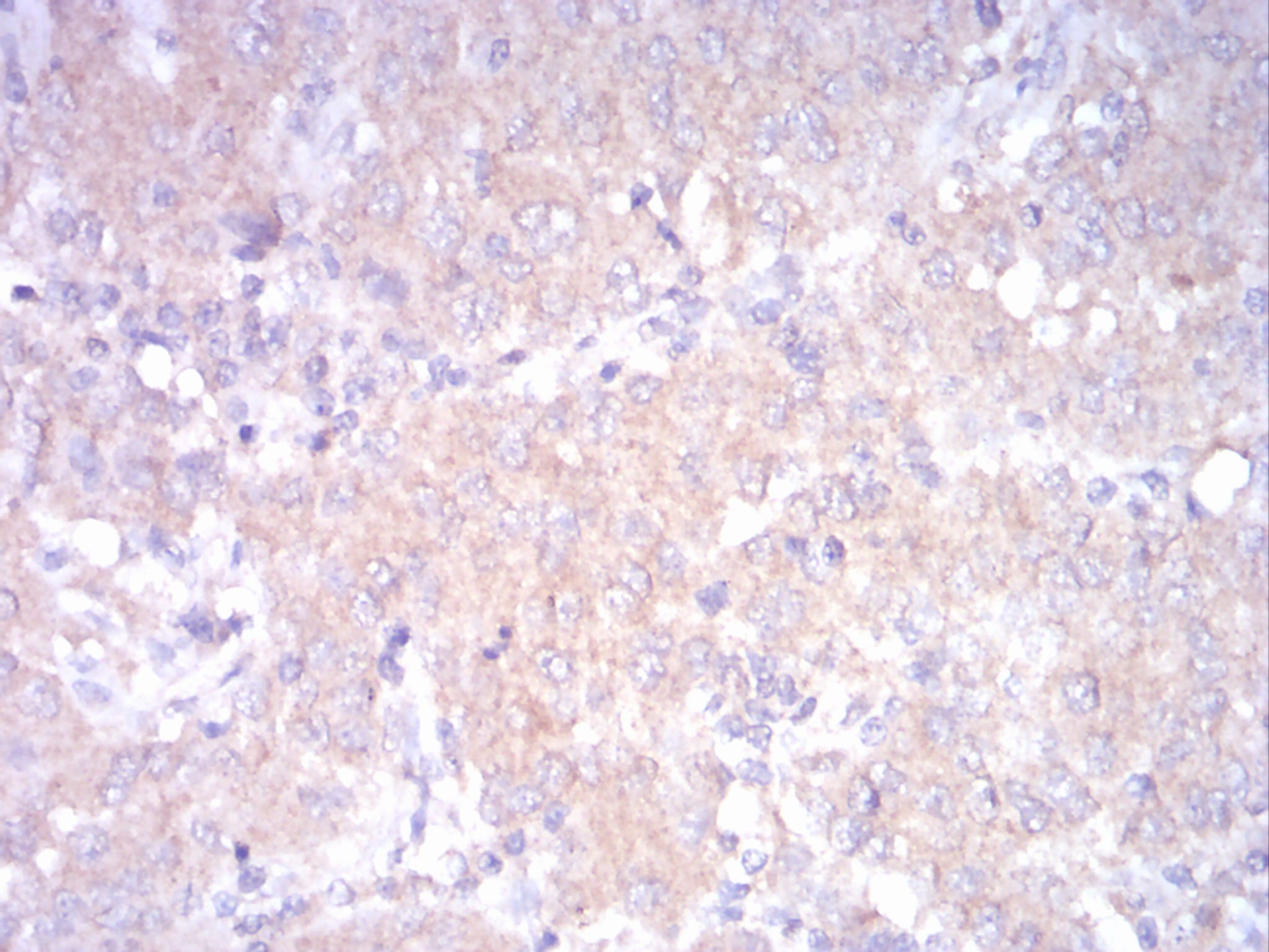
Figure 5:Immunohistochemical analysis of paraffin-embedded ovarian cancertissues using LDLR mouse mAb with DAB staining.
For Research Use Only. Not for use in diagnostic procedures.