

KRT19 Primary Antibody
Item Information
Catalog #
Size
Price
Description
The protein encoded by this gene is a member of the keratin family. The keratins are intermediate filament proteins responsible for the structural integrity of epithelial cells and are subdivided into cytokeratins and hair keratins. The type I cytokeratins consist of acidic proteins which are arranged in pairs of heterotypic keratin chains. Unlike its related family members, this smallest known acidic cytokeratin is not paired with a basic cytokeratin in epithelial cells. It is specifically expressed in the periderm, the transiently superficial layer that envelopes the developing epidermis. The type I cytokeratins are clustered in a region of chromosome 17q12-q21.
Product Overview
Entrez GenelD
3880
Aliases
K19; CK19; K1CS; MGC15366
Clone#
4E8
Host / Isotype
Mouse / IgG1
Species Reactivity
Human
Immunogen
Purified recombinant fragment of human KRT19 expressed in E. Coli.
Formulation
Ascitic fluid containing 0.03% sodium azide.
Storage
Store at 4°C short term. Aliquot and store at -20°C long term. Avoid freeze/thaw cycles.
Product Applications
WB (Western Blot)
1/500 - 1/2000
IHC_P(Immunohistochemistry)
1/200 - 1/1000
ICC (Immunocytochemistry)
1/200 - 1/1000
ELISA
1/10000
References
1. Exp Cell Res. 2009 Jul 1;315(11):1964-74.
2. Acta Cytol. 2008 Sep-Oct;52(5):541-8.
2. Acta Cytol. 2008 Sep-Oct;52(5):541-8.
Product Image
Western Blot

Figure 1: Western blot analysis using KRT19 mAb against human KRT19 (AA: 115-269) recombinant protein. (Expected MW is 43.1 kDa)
Western Blot
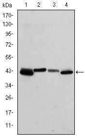
Figure 2: Western blot analysis using KRT19 mouse mAb against T47D (1), MCF-7 (2), HepG2 (3) and SW620 (4) cell lysate.
Immunohistochemical analysis

Figure 3: Immunohistochemical analysis of paraffin-embedded human cervical cancer tissues using KRT19 mouse mAb with DAB staining.
Immunohistochemical analysis

Figure 4: Immunohistochemical analysis of paraffin-embedded human colon cancer tissues using KRT19 mouse mAb with DAB staining.
Immunohistochemical analysis

Figure 5: Immunohistochemical analysis of paraffin-embedded human stomach cancer tissues using KRT19 mouse mAb with DAB staining.
Immunohistochemical analysis

Figure 6: Immunohistochemical analysis of paraffin-embedded human bladder cancer tissues using KRT19 mouse mAb with DAB staining.
Immunofluorescence analysis

Figure 7: Immunofluorescence analysis of HepG2 cells using KRT19 mouse mAb (green). Blue: DRAQ5 fluorescent DNA dye. Red: Actin filaments have been labeled with Alexa Fluor-555 phalloidin.
Elisa

Red: Control Antigen (100ng); Purple: Antigen (10ng); Green: Antigen (50ng); Blue: Antigen (100ng);
For Research Use Only. Not for use in diagnostic procedures.