

KRT18 Primary Antibody
Item Information
Catalog #
Size
Price
Description
KRT18 encodes the type I intermediate filament chain keratin 18. Keratin 18, together with its filament partner keratin 8, are perhaps the most commonly found members of the intermediate filament gene family. They are expressed in single layer epithelial tissues of the body. Mutations in this gene have been linked to cryptogenic cirrhosis. Two transcript variants encoding the same protein have been found for this gene.
Product Overview
Entrez GenelD
3875
Aliases
K18; CK-18; CYK18
Clone#
3G6E4
Host / Isotype
Mouse / Mouse IgG1
Immunogen
Purified recombinant fragment of human KRT18 (AA: 1-150) expressed in E. Coli.
Formulation
Purified antibody in PBS with 0.05% sodium azide
Storage
Store at 4°C short term. Aliquot and store at -20°C long term. Avoid freeze/thaw cycles.
Product Applications
WB (Western Blot)
1/500 - 1/2000
IHC_P(Immunohistochemistry)
1/200 - 1/1000
ICC (Immunocytochemistry)
1/500 - 1/2000
FCM (Flow Cytometry)
1/200 - 1/400
ELISA
1/10000
References
1.Leukemia. 2018 Dec;32(12):2685-2692. 2.BMC Neurol. 2018 Mar 24;18(1):32.
Product Image
Elisa

Figure 1:Black line: Control Antigen (100 ng);Purple line: Antigen (10ng); Blue line: Antigen (50 ng); Red line:Antigen (100 ng)
Western Blot

Figure 2:Western blot analysis using KRT18 mAb against human KRT18 (AA: 1-150) recombinant protein. (Expected MW is 42.1 kDa)
Western Blot

Figure 3:Western blot analysis using KRT18 mAb against HEK293 (1) and KRT18 (AA: 1-150)-hIgGFc transfected HEK293 (2) cell lysate.
Western Blot

Figure 4:Western blot analysis using KRT18 mouse mAb against SK-BR-3 (1), HepG2 (2), T47D (3), MCF-7 (4), Hela (5), A431 (6), and HCT116 (7) cell lysate.
Immunofluorescence analysis

Figure 5:Immunofluorescence analysis of Hela cells using KRT18 mouse mAb (green). Blue: DRAQ5 fluorescent DNA dye. Red: Actin filaments have been labeled with Alexa Fluor- 555 phalloidin. Secondary antibody from Fisher (Cat#: 35503)
Flow Cytometric
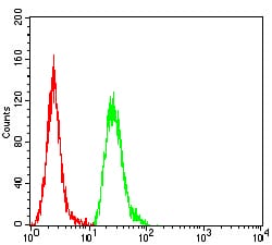
Figure 6:Flow cytometric analysis of Hela cells using KRT18 mouse mAb (green) and negative control (red).
Immunohistochemical Analysis

Figure 7:Immunohistochemical analysis of paraffin-embedded lung cancer tissues using KRT18 mouse mAb with DAB staining.
Immunohistochemical Analysis

Figure 8:Immunohistochemical analysis of paraffin-embedded rectum cancer tissues using KRT18 mouse mAb with DAB staining.
For Research Use Only. Not for use in diagnostic procedures.