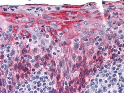
KRT15 Primary Antibody

Item Information
Catalog #
Size
Price
Description

The protein encoded by this gene is a member of the keratin gene family. The keratins are intermediate filament proteins responsible for the structural integrity of epithelial cells and are subdivided into cytokeratins and hair keratins. Keratin 15 is a type I keratin without a defined type II partner. Keratin 15 is expressed primarily in the basal keratinocytes of stratified tissues, including the fetal epidermis and fetal nail. Expression of keratin 15 is downregulated in some hyperproliferating situations, such as psoriasis and hypertrophic scars. Because keratinocytes in psoriasis and hypertrophic scars are activated, it is suggested that keratin 15 expression is not compatible with keratinocyte activation and the keratin 15 gene is downregulated to maintain the activated phenotype.

Product Overview
Entrez GenelD
3866
Aliases
K15; CK15; K1CO; KRT1
Clone#
6E7
Host / Isotype
Mouse / IgG2a
Species Reactivity
Human
Immunogen
Purified recombinant fragment of KRT15 expressed in E. Coli.
Formulation
Ascitic fluid containing 0.03% sodium azide.
Storage
4°C; -20°C for long term storage
Product Applications
WB (Western Blot)
1/500 - 1/2000
IHC_P(Immunohistochemistry)
1/200 - 1/1000
ICC (Immunocytochemistry)
1/200 - 1/1000
FCM (Flow Cytometry)
1/200 - 1/400
ELISA
1/10000
References
1. J Invest Dermatol. 1999. 112(3):362-9
2. Exp Cell Res. 2006. 254(1):80-90
3. Mol Cell Biol. 2000. 24(8):3168-79
Product Image
Western Blot
Figure 1: Western blot analysis using KRT15 mouse mAb against A431 cell lysate.
Immunohistochemical analysis
Figure 2: Immunohistochemical analysis of paraffin-embedded human Tonsil tissues using KRT15 mouse mAb
Immunofluorescence analysis
Figure 3: Immunofluorescence analysis of HepG2(left) and PACN-1 (right) cells using KRT15 mouse mAb (green). Red: Actin filaments have been labeled with DY-554 phalloidin. Blue: DRAQ5 fluorescent DNA dye.
Flow cytometric
Figure 4: Flow cytometric analysis of PACN-1 cells using KRT15 mouse mAb (green) and negative control (purple).
For Research Use Only. Not for use in diagnostic procedures.