

Mouse Monoclonal Antibody to KRT10
Item Information
Catalog #
Size
Price
Description
This gene encodes a member of the type I (acidic) cytokeratin family, which belongs to the superfamily of intermediate filament (IF) proteins. Keratins are heteropolymeric structural proteins which form the intermediate filament. These filaments, along with actin microfilaments and microtubules, compose the cytoskeleton of epithelial cells. Mutations in this gene are associated with epidermolytic hyperkeratosis. This gene is located within a cluster of keratin family members on chromosome 17q21.
Product Overview
Entrez GenelD
3858
Aliases
BIE; EHK; K10; KPP; BCIE; CK10
Clone#
7A5A6
Host / Isotype
Mouse / IgG1
Immunogen
Purified recombinant fragment of human KRT10 (AA: 146-455) expressed in E. Coli.
Formulation
Purified antibody in PBS with 0.05% sodium azide
Storage
Store at 4°C short term. Aliquot and store at -20°C long term. Avoid freeze/thaw cycles.
Product Applications
WB (Western Blot)
1/500 - 1/2000
IHC_P(Immunohistochemistry)
1/200 - 1/1000
ICC (Immunocytochemistry)
1/200 - 1/1000
FCM (Flow Cytometry)
1/200 - 1/400
ELISA
1/10000
References
1.J Cell Mol Med. 2019 Dec;23(12):8442-8452. 2.J Eur Acad Dermatol Venereol. 2016 Oct;30(10):e102-e104.
Product Image
Elisa
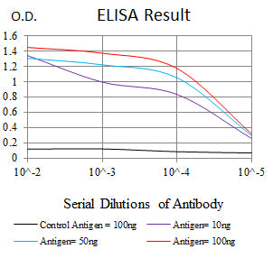
Figure 1:Black line: Control Antigen (100 ng);Purple line: Antigen (10ng); Blue line: Antigen (50 ng); Red line:Antigen (100 ng)
Western Blot
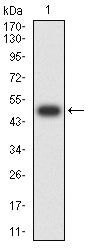
Figure 2:Western blot analysis using KRT10 mAb against human KRT10 (AA: 146-455) recombinant protein. (Expected MW is 49.1 kDa)
Western Blot
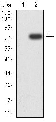
Figure 3:Western blot analysis using KRT10 mAb against HEK293-6e (1) and KRT10 (AA: 146-455)-hIgGFc transfected HEK293-6e (2) cell lysate.
Western Blot
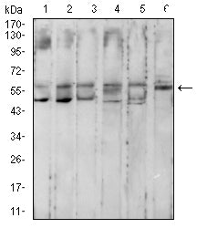
Figure 4:Western blot analysis using KRT10 mouse mAb against MCF-7 (1), Hela (2), HepG2 (3), T47D (4), HT-29 (5), and A549 (6) cell lysate.
Immunofluorescence analysis
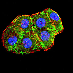
Figure 5:Immunofluorescence analysis of Hela cells using KRT10 mouse mAb (green). Blue: DRAQ5 fluorescent DNA dye. Red: Actin filaments have been labeled with Alexa Fluor- 555 phalloidin. Secondary antibody from Fisher (Cat#: 35503)
Flow cytometric analysis
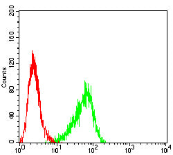
Figure 6:Flow cytometric analysis of A431 cells using KRT10 mouse mAb (green) and negative control (red).
Immunohistochemical analysis
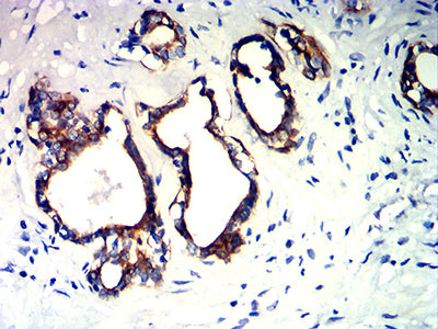
Figure 7:Immunohistochemical analysis of paraffin-embedded prostate cancer tissues using KRT10 mouse mAb with DAB staining.
Immunohistochemical analysis
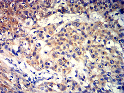
Figure 8:Immunohistochemical analysis of paraffin-embedded esophageal cancer tissues using KRT10 mouse mAb with DAB staining.
For Research Use Only. Not for use in diagnostic procedures.