

KEAP1 Primary Antibody
Item Information
Catalog #
Size
Price
Description
This gene encodes a protein containing KELCH-1 like domains, as well as a BTB/POZ domain. Kelch-like ECH-associated protein 1 interacts with NF-E2-related factor 2 in a redox-sensitive manner and the dissociation of the proteins in the cytoplasm is followed by transportation of NF-E2-related factor 2 to the nucleus. This interaction results in the expression of the catalytic subunit of gamma-glutamylcysteine synthetase. Two alternatively spliced transcript variants encoding the same isoform have been found for this gene.
Product Overview
Entrez GenelD
9817
Aliases
INrf2; KLHL19
Clone#
1F10B6
Host / Isotype
Mouse / IgG1
Species Reactivity
Human, Mouse
Immunogen
Purified recombinant fragment of human KEAP1 (AA: 380-624) expressed in E. Coli.
Formulation
Ascitic fluid containing 0.03% sodium azide.
Storage
Store at 4°C short term. Aliquot and store at -20°C long term. Avoid freeze/thaw cycles.
Product Applications
WB (Western Blot)
1/500 - 1/2000
ICC (Immunocytochemistry)
1/50
FCM (Flow Cytometry)
1/200 - 1/400
ELISA
1/10000
References
1.Cell Signal. 2010 Nov;22(11):1645-54.
2.Mol Cancer Ther. 2010 Feb;9(2):336-46.
2.Mol Cancer Ther. 2010 Feb;9(2):336-46.
Product Image
Western Blot

Figure 1: Western blot analysis using KEAP1 mAb against human KEAP1 recombinant protein. (Expected MW is 52.7 kDa)
Western Blot

Figure 2: Western blot analysis using KEAP1 mouse mAb against NIH3T3 (1), and A549 (2) cell lysate.
Immunofluorescence analysis
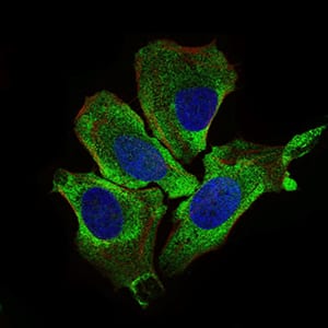
Figure 3: Immunofluorescence analysis of HeLa cells using KEAP1 mouse mAb (green). Blue: DRAQ5 fluorescent DNA dye. Red: Actin filaments have been labeled with Alexa Fluor-555 phalloidin.
Flow cytometric

Figure 4: Flow cytometric analysis of HepG2 cells using KEAP1 mouse mAb (green) and negative control (purple).
Elisa

Black line: Control Antigen (100 ng); Purple line: Antigen(10ng); Blue line: Antigen (50 ng); Red line: Antigen (100 ng);
For Research Use Only. Not for use in diagnostic procedures.