

KDM1A Primary Antibody
Item Information
Catalog #
Size
Price
Description
This gene encodes a nuclear protein containing a SWIRM domain, a FAD-binding motif, and an amine oxidase domain. This protein is a component of several histone deacetylase complexes, though it silences genes by functioning as a histone demethylase. Alternative splicing results in multiple transcript variants.
Product Overview
Entrez GenelD
23028
Aliases
AOF2; CPRF; KDM1; LSD1; BHC110
Clone#
3F9A3
Host / Isotype
Mouse / IgG1
Species Reactivity
Human, Monkey
Immunogen
Purified recombinant fragment of human KDM1A (AA: 55-263) expressed in E. Coli.
Formulation
Purified antibody in PBS with 0.05% sodium azide
Storage
Store at 4°C short term. Aliquot and store at -20°C long term. Avoid freeze/thaw cycles.
Product Applications
WB (Western Blot)
1/500 - 1/2000
IHC_P(Immunohistochemistry)
1/200 - 1/1000
ICC (Immunocytochemistry)
1/200 - 1/1000
FCM (Flow Cytometry)
1/200 - 1/400
ELISA
1/10000
References
1.Int J Clin Exp Pathol. 2014 Dec 1;7(12):8929-34.
2.Blood. 2014 Jul 3;124(1):151-2.
2.Blood. 2014 Jul 3;124(1):151-2.
Product Image
Elisa
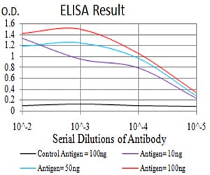
Figure 1: Black line: Control Antigen (100 ng);Purple line: Antigen (10ng); Blue line: Antigen (50 ng); Red line:Antigen (100 ng)
Western Blot
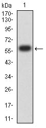
Figure 2:Western blot analysis using KDM1A mAb against human KDM1A (AA: 55-263) recombinant protein. (Expected MW is 60.3 kDa)
Western Blot
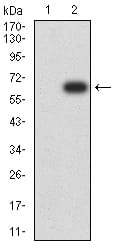
Figure 3:Western blot analysis using KDM1A mAb against HEK293 (1) and KDM1A (AA: 55-263)-hIgGFc transfected HEK293 (2) cell lysate.
Western Blot
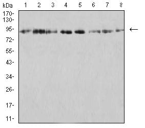
Figure 4:Western blot analysis using KDM1A mouse mAb against SK-Br-3 (1), K562 (2), SW480 (3), Jurkat (4), Hela (5), COS7 (6), T47D (7), and HCT116 (8) cell lysate.
Flow cytometric
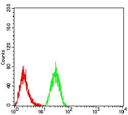
Figure 5:Flow cytometric analysis of Hela cells using KDM1A mouse mAb (green) and negative control (red).
Immunofluorescence analysis
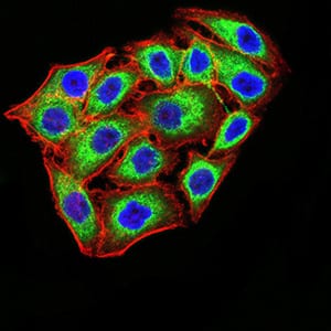
Figure 6:Immunofluorescence analysis of MCF-7 cells using KDM1A mouse mAb (green). Blue: DRAQ5 fluorescent DNA dye. Red: Actin filaments have been labeled with Alexa Fluor- 555 phalloidin. Secondary antibody from Fisher (Cat#: 35503)
Immunohistochemical analysis

Figure 7:Immunohistochemical analysis of paraffin-embedded colon cancer tissues using KDM1A mouse mAb with DAB staining.
Immunohistochemical analysis
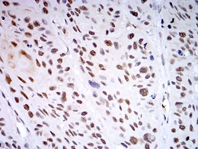
Figure 8:Immunohistochemical analysis of paraffin-embedded esophageal cancer tissues using KDM1A mouse mAb with DAB staining.
For Research Use Only. Not for use in diagnostic procedures.