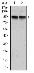
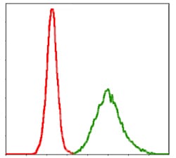
KCNQ1 Primary Antibody

Item Information
Catalog #
Size
Price
Description

This gene encodes a voltage-gated potassium channel required for repolarization phase of the cardiac action potential. This protein can form heteromultimers with two other potassium channel proteins, KCNE1 and KCNE3. Mutations in this gene are associated with hereditary long QT syndrome 1 (also known as Romano-Ward syndrome), Jervell and Lange-Nielsen syndrome, and familial atrial fibrillation. This gene exhibits tissue-specific imprinting, with preferential expression from the maternal allele in some tissues, and biallelic expression in others. This gene is located in a region of chromosome 11 amongst other imprinted genes that are associated with Beckwith-Wiedemann syndrome (BWS), and itself has been shown to be disrupted by chromosomal rearrangements in patients with BWS. Alternatively spliced transcript variants have been found for this gene. 

Product Overview
Entrez GenelD
3784
Aliases
LQT; RWS; WRS; LQT1; SQT2; ATFB1; ATFB3; JLNS1; KCNA8; KCNA9; Kv1.9; Kv7.1; KVLQT1; FLJ26167
Clone#
5E12
Host / Isotype
Mouse / IgG2b
Species Reactivity
Human
Immunogen
Purified recombinant fragment of human KCNQ1 expressed in E. Coli.
Formulation
Ascitic fluid containing 0.03% sodium azide.
Storage
4°C; -20°C for long term storage
Product Applications
WB (Western Blot)
1/500 - 1/2000
FCM (Flow Cytometry)
1/200 - 1/400
ELISA
1/10000
References
1. Biochem Biophys Res Commun. 2009 May 29;383(2):206-9. 
2. J Biol Chem. 2009 Jun 12;284(24):16452-62. 
Product Image
Western Blot
Figure 1: Western blot analysis using KCNQ1 mAb against human KCNQ1 (AA: 229-347) recombinant protein. (Expected MW is 74.7 kDa)
Western Blot
Figure 2: Western blot analysis using KCNQ1 mouse mAb against MCF-7 (1) and A431 (2) cell lysate.
Flow cytometric
Figure 3: Flow cytometric analysis of MCF-7 cells using KCNQ1 mouse mAb (green) and negative control (red).
Elisa
Black line: Control Antigen (100 ng); Purple line: Antigen(10ng); Blue line: Antigen (50 ng); Red line: Antigen (100 ng);
For Research Use Only. Not for use in diagnostic procedures.