

JAK2 Primary Antibody
Item Information
Catalog #
Size
Price
Description
This gene product is a protein tyrosine kinase involved in a specific subset of cytokine receptor signaling pathways. It has been found to be constituitively associated with the prolactin receptor and is required for responses to gamma interferon. Mice that do not express an active protein for this gene exhibit embryonic lethality associated with the absence of definitive erythropoiesis.
Product Overview
Entrez GenelD
3717
Aliases
JTK10; THCYT3
Clone#
5C12C9
Host / Isotype
Mouse / IgG1
Species Reactivity
Human
Immunogen
Purified recombinant fragment of human JAK2 (AA: 745-955) expressed in E. Coli.
Formulation
Purified antibody from tissue culture in PBS with 0.05% sodium azide
Storage
Store at 4°C short term. Aliquot and store at -20°C long term. Avoid freeze/thaw cycles.
Product Applications
WB (Western Blot)
1/500 - 1/2000
IHC_P(Immunohistochemistry)
1/200 - 1/1000
FCM (Flow Cytometry)
1/200 - 1/400
ELISA
1/10000
References
Blood. 2014 May 1;123(18):2826-37.
PLoS One. 2013 May 24;8(5):e64628.
PLoS One. 2013 May 24;8(5):e64628.
Product Image
Western Blot
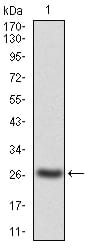
Figure 1: Western blot analysis using JAK2 mAb against human JAK2(AA: 745-955) recombinant protein. (Expected MW is 27.1 kDa)
Western Blot
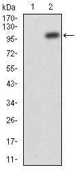
Figure 2: Western blot analysis using JAK2 mAb against HEK293 (1) and JAK2(AA: 545-1124)-hIgGFc transfected HEK293 (2) cell lysate.
Flow cytometric
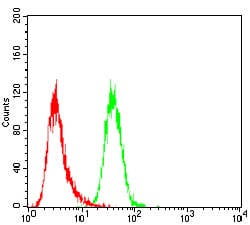
Figure 3: Flow cytometric analysis of Hela cells using JAK2 mouse mAb (green) and negative control (red).
Immunohistochemical analysis
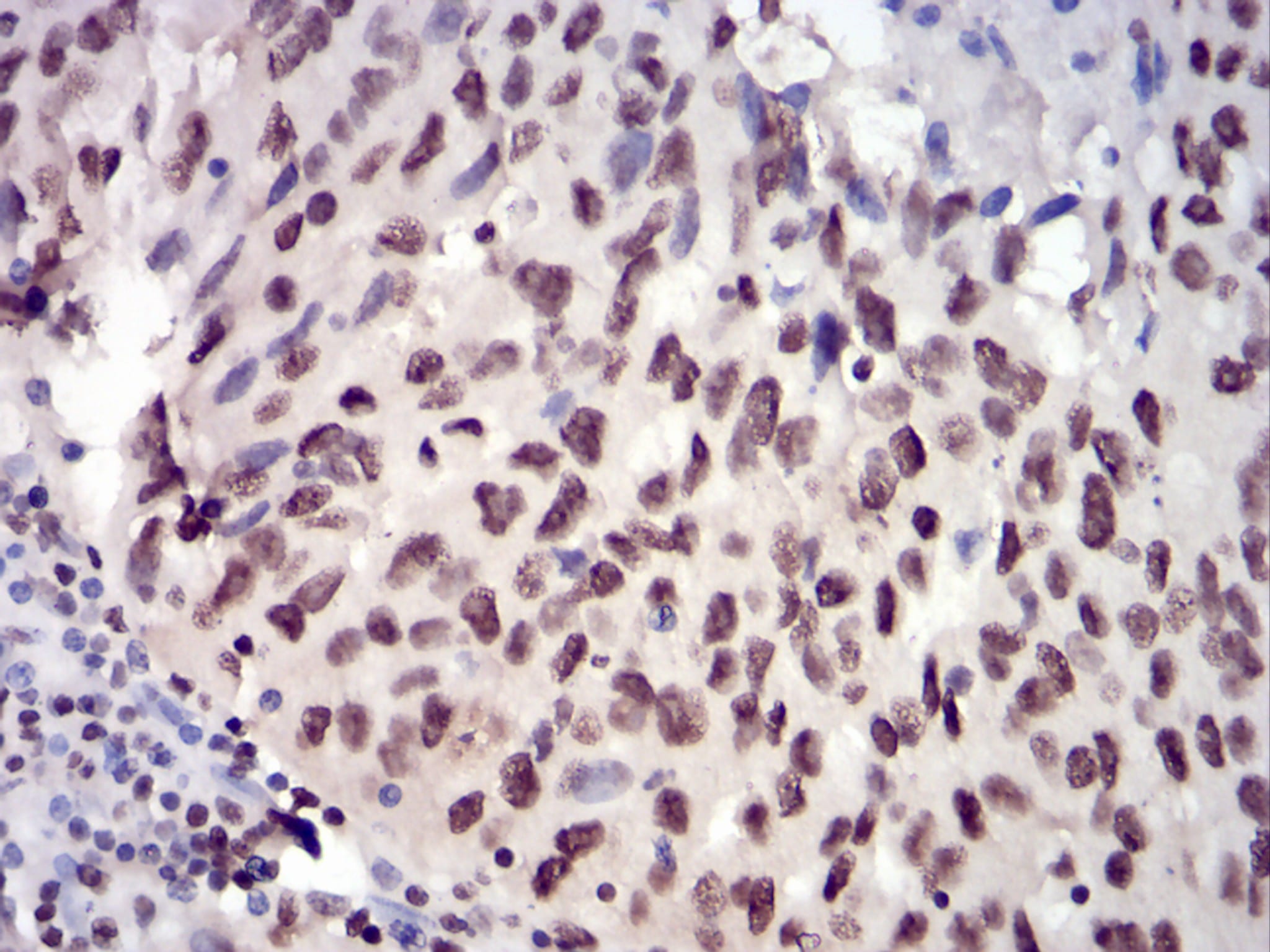
Figure 4: Immunohistochemical analysis of paraffin-embedded ovarian cancer tissues using JAK2 mouse mAb with DAB staining.
Immunohistochemical analysis

Figure 5: Immunohistochemical analysis of paraffin-embedded breast cancer tissues using JAK2 mouse mAb with DAB staining.
Elisa
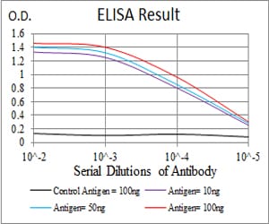
Black line: Control Antigen (100 ng); Purple line: Antigen(10ng); Blue line: Antigen (50 ng); Red line: Antigen (100 ng);
For Research Use Only. Not for use in diagnostic procedures.