
Mouse Monoclonal Antibody to ITGA1
Item Information
Catalog #
Size
Price
Description
This gene encodes the alpha 1 subunit of integrin receptors. This protein heterodimerizes with the beta 1 subunit to form a cell-surface receptor for collagen and laminin. The heterodimeric receptor is involved in cell-cell adhesion and may play a role in inflammation and fibrosis. The alpha 1 subunit contains an inserted (I) von Willebrand factor type I domain which is thought to be involved in collagen binding. [provided by RefSeq, Jul 2008]
Product Overview
Entrez GenelD
3672
Aliases
VLA1; CD49a
Clone#
5F9H10
Host / Isotype
Mouse / IgG1
Immunogen
Purified recombinant fragment of humanITGA1 (AA: extra(151-364)) expressed in E. Coli.
Formulation
Purified antibody in PBS with 0.05% sodium azide
Storage
Store at 4°C short term. Aliquot and store at -20°C long term. Avoid freeze/thaw cycles.
Product Applications
WB (Western Blot)
1/500 - 1/2000
IHC_P(Immunohistochemistry)
1/200 - 1/1000
FCM (Flow Cytometry)
1/200 - 1/400
ELISA
1/10000
References
1.Sci Rep.2017 Aug 30;7(1):10060.2.Structure.2018 Aug 7;26(8):1080-1090.e5.
Product Image
Elisa

Figure 1:Black line: Control Antigen (100 ng);Purple line: Antigen (10ng); Blue line: Antigen (50 ng); Red line:Antigen (100 ng)
Western Blot

Figure 2:Western blot analysis using ITGA1 mAb against human ITGA1 (AA: extra(151-364)) recombinant protein. (Expected MW is 26.7 kDa)
Western Blot

Figure 3:Western blot analysis using ITGA1 mAb against HEK293 (1) and ITGA1 (AA: extra(151-364))-hIgGFc transfected HEK293--6e (2) cell lysate.
Flow cytometric analysis

Figure 4:Flow cytometric analysis of Hela cells using ITGA1 mouse mAb (green) and negative control (red).
Flow cytometric analysis

Figure 5:Flow cytometric analysis of HrpG2 cells using ITGA1 mouse mAb (green) and negative control (red).
Immunohistochemical analysis
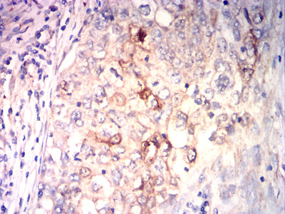
Figure 6:Immunohistochemical analysis of paraffin-embedded Lung cancer tissues using ITGA1 mouse mAb with DAB staining.
Immunohistochemical analysis

Figure 7:Immunohistochemical analysis of paraffin-embedded Prostate cancer tissues using ITGA1 mouse mAb with DAB staining.
For Research Use Only. Not for use in diagnostic procedures.