

IRF3 Primary Antibody
Item Information
Catalog #
Size
Price
Description
This gene encodes a member of the interferon regulatory transcription factor (IRF) family. The encoded protein is found in an inactive cytoplasmic form that upon serine/threonine phosphorylation forms a complex with CREBBP. This complex translocates to the nucleus and activates the transcription of interferons alpha and beta, as well as other interferon-induced genes. The protein plays an important role in the innate immune response against DNA and RNA viruses. Mutations in this gene are associated with Encephalopathy, acute, infection-induced, herpes-specific, 7.
Product Overview
Entrez GenelD
3661
Aliases
IIAE7
Clone#
5G3E2
Host / Isotype
Mouse / Mouse IgG1
Species Reactivity
Human, Mouse, Rat
Immunogen
Purified recombinant fragment of human IRF3 (AA: 1-150) expressed in HEK293-6e cells supernatant.
Formulation
Purified antibody in PBS with 0.05% sodium azide
Storage
Store at 4°C short term. Aliquot and store at -20°C long term. Avoid freeze/thaw cycles.
Product Applications
WB (Western Blot)
1/500 - 1/2000
IHC_P(Immunohistochemistry)
1/200 - 1/1000
FCM (Flow Cytometry)
1/200 - 1/400
ELISA
1/10000
References
1.J Immunol. 2020 Oct 15;205(8):1981-1989.
2.Int J Biol Sci. 2021 Apr 10;17(6):1547-1554.
2.Int J Biol Sci. 2021 Apr 10;17(6):1547-1554.
Product Image
Elisa

Figure 1:Black line: Control Antigen (100 ng);Purple line: Antigen (10ng); Blue line: Antigen (50 ng); Red line:Antigen (100 ng)
Western Blot

Figure 2:Western blot analysis using IRF3 mAb against human IRF3 (AA: 1-150) recombinant protein. (Expected MW is 47.4 kDa)
Western Blot

Figure 3:Western blot analysis using IRF3 mouse mAb against THP-1 (1), Hela (2), A549 (3), MCF-7 (4), K562 (5), Jurkat (6) and A431 (7) cell lysate.
Immunofluorescence analysis

Figure 4:Flow cytometric analysis of Hela cells using IRF3 mouse mAb (green) and negative control (red).
Immunohistochemical analysis

Figure 5:Immunohistochemical analysis of paraffin-embedded human bladder cancer tissues using IRF3 mouse mAb with DAB staining.
Immunohistochemical analysis

Figure 6:Immunohistochemical analysis of paraffin-embedded human colon cancer tissues using IRF3 mouse mAb with DAB staining.
Immunohistochemical analysis
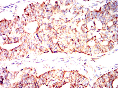
Figure 7:Immunohistochemical analysis of paraffin-embedded human ovarian cancer tissues using IRF3 mouse mAb with DAB staining.
Immunohistochemical analysis

Figure 8:Immunohistochemical analysis of paraffin-embedded human rectum cancer tissues using IRF3 mouse mAb with DAB staining.
Immunohistochemical analysis

Figure 9:Immunohistochemical analysis of paraffin-embedded mouse kidney tissues using IRF3 mouse mAb with DAB staining.
Immunohistochemical analysis

Figure 10:Immunohistochemical analysis of paraffin-embedded mouse brain tissues using IRF3 mouse mAb with DAB staining.
Immunohistochemical analysis

Figure 11:Immunohistochemical analysis of paraffin-embedded rat kidney tissues using IRF3 mouse mAb with DAB staining.
Immunohistochemical analysis

Figure 12:Immunohistochemical analysis of paraffin-embedded rat brain tissues using IRF3 mouse mAb with DAB staining.
Immunohistochemical analysis

Figure 13:Immunohistochemical analysis of paraffin-embedded rabbit kidney tissues using IRF3 mouse mAb with DAB staining.
Immunohistochemical analysis

Figure 14:Immunohistochemical analysis of paraffin-embedded rabbit brain tissues using IRF3 mouse mAb with DAB staining.
For Research Use Only. Not for use in diagnostic procedures.