

IRAK4 Primary Antibody
Item Information
Catalog #
Size
Price
Description
This gene encodes a kinase that activates NF-kappaB in both the Toll-like receptor (TLR) and T-cell receptor (TCR) signaling pathways. The protein is essential for most innate immune responses. Mutations in this gene result in IRAK4 deficiency and recurrent invasive pneumococcal disease. Multiple transcript variants encoding different isoforms have been found for this gene.
Product Overview
Entrez GenelD
51135
Aliases
IPD1; REN64; NY-REN-64
Clone#
2H9
Host / Isotype
Mouse / IgG1
Species Reactivity
Human, Mouse, Monkey
Immunogen
Purified recombinant fragment of human IRAK4 expressed in E. Coli.
Formulation
Ascitic fluid containing 0.03% sodium azide.
Storage
Store at 4°C short term. Aliquot and store at -20°C long term. Avoid freeze/thaw cycles.
Product Applications
WB (Western Blot)
1/500 - 1/2000
IHC_P(Immunohistochemistry)
1/200 - 1/1000
FCM (Flow Cytometry)
1/200 - 1/400
ELISA
1/10000
References
1. J Biol Chem. 2010 Jun 11;285(24):18276-82.
2. Scand J Immunol. 2009 Sep;70(3):264-76.
2. Scand J Immunol. 2009 Sep;70(3):264-76.
Product Image
Western Blot

Figure 1: Western blot analysis using IRAK4 mAb against human IRAK4 (AA: 21-198) recombinant protein. (Expected MW is 45.4 kDa)
Western Blot

Figure 2: Western blot analysis using IRAK4 mouse mAb against THP-1 (1), Hela (2), K562 (3), MCF-7 (4), RAW264.7 (5), Jurkat (6) and Cos7 (7) cell lysate.
Immunohistochemical analysis

Figure 3: Immunohistochemical analysis of paraffin-embedded human lung cancer tissues using IRAK4 mouse mAb with DAB staining.
Immunohistochemical analysis

Figure 4: Immunohistochemical analysis of paraffin-embedded human kidney cancer tissues using IRAK4 mouse mAb with DAB staining.
Flow cytometric

Figure 5: Flow cytometric analysis of Hela cells using IRAK4 mouse mAb (blue) and negative control (red).
Elisa
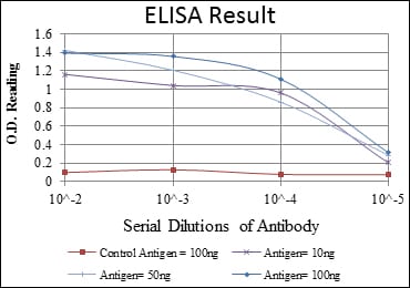
Red: Control Antigen (100ng); Purple: Antigen (10ng); Green: Antigen (50ng); Blue: Antigen (100ng);
For Research Use Only. Not for use in diagnostic procedures.