

IL2RA Primary Antibody
Item Information
Catalog #
Size
Price
Description
The interleukin 2 (IL2) receptor alpha (IL2RA) and beta (IL2RB) chains, together with the common gamma chain (IL2RG), constitute the high-affinity IL2 receptor. Homodimeric alpha chains (IL2RA) result in low-affinity receptor, while homodimeric beta (IL2RB) chains produce a medium-affinity receptor. Normally an integral-membrane protein, soluble IL2RA has been isolated and determined to result from extracellular proteolyisis. Alternately-spliced IL2RA mRNAs have been isolated, but the significance of each is presently unknown. Mutations in this gene are associated with interleukin 2 receptor alpha deficiency.
Product Overview
Entrez GenelD
3559
Aliases
CD25; IL2R; TCGFR; IDDM10
Clone#
1B5D12
Host / Isotype
Mouse / IgG1
Species Reactivity
Human, Mouse, Monkey, Rat
Immunogen
Purified recombinant fragment of human IL2RA (AA: 34-139) expressed in E. Coli.
Formulation
Purified antibody in PBS with 0.05% sodium azide
Storage
Store at 4°C short term. Aliquot and store at -20°C long term. Avoid freeze/thaw cycles.
Product Applications
WB (Western Blot)
1/500 - 1/2000
IHC_P(Immunohistochemistry)
1/200 - 1/1000
ICC (Immunocytochemistry)
1/200 - 1/1000
ELISA
1/10000
References
1. Ann Hematol. 2012 Oct;91(10):1597-602.
2. Transplant Proc. 2012 May;44(4):1139-42.
2. Transplant Proc. 2012 May;44(4):1139-42.
Product Image
Western Blot

Figure 1: Western blot analysis using IL2RA mAb against human IL2RA recombinant protein. (Expected MW is 37.5 kDa)
Western Blot
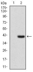
Figure 2: Western blot analysis using IL2RA mAb against HEK293 (1) and IL2RA (AA: 34-139)-hIgGFc transfected HEK293 (2) cell lysate.
Immunofluorescence analysis

Figure 3: Immunofluorescence analysis of Hela cells using IL2RA mouse mAb (green). Blue: DRAQ5 fluorescent DNA dye.
Western Blot

Figure 3: Western blot analysis using IL2RA mouse mAb against Hela (1), MOLT4 (2), HEK293 (3), A549 (4), Jurkat (5), K562 (6), Cos7 (7), PC-12 (8) and NIH/3T3 (9) cell lysate.
Immunohistochemical analysis

Figure 4: Immunohistochemical analysis of paraffin-embedded rectum cancer tissues using IL2RA mouse mAb with DAB staining.
Immunohistochemical analysis

Figure 5: Immunohistochemical analysis of paraffin-embedded bladder cancer tissues using IL2RA mouse mAb with DAB staining.
Elisa

Black line: Control Antigen (100 ng); Purple line: Antigen(10ng); Blue line: Antigen (50 ng); Red line: Antigen (100 ng);
For Research Use Only. Not for use in diagnostic procedures.