

IL28A Primary Antibody
Item Information
Catalog #
Size
Price
Description
This gene encodes a cytokine distantly related to type I interferons and the IL-10 family. This gene, interleukin 28B (IL28B), and interleukin 29 (IL29) are three closely related cytokine genes that form a cytokine gene cluster on a chromosomal region mapped to 19q13. Expression of the cytokines encoded by the three genes can be induced by viral infection. All three cytokines have been shown to interact with a heterodimeric class II cytokine receptor that consists of interleukin 10 receptor, beta (IL10RB) and interleukin 28 receptor, alpha (IL28RA).
Product Overview
Entrez GenelD
282616
Aliases
IFNL2; IL-28A
Clone#
6H9E6
Host / Isotype
Mouse / IgG1
Species Reactivity
Human
Immunogen
Purified recombinant fragment of human IL28A (AA: 1-200) expressed in E. Coli.
Formulation
Purified antibody in PBS with 0.05% sodium azide
Storage
Store at 4°C short term. Aliquot and store at -20°C long term. Avoid freeze/thaw cycles.
Product Applications
WB (Western Blot)
1/500 - 1/2000
ICC (Immunocytochemistry)
1/200 - 1/1000
ELISA
1/10000
References
1.Cell Immunol. 2014 Jul;290(1):116-9.
2.Cell Signal. 2012 Sep;24(9):1734-42.
2.Cell Signal. 2012 Sep;24(9):1734-42.
Product Image
Elisa

Figure 1: Black line: Control Antigen (100 ng);Purple line: Antigen (10ng); Blue line: Antigen (50 ng); Red line:Antigen (100 ng)
Western Blot
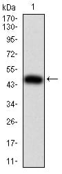
Figure 2:Western blot analysis using IL28A mAb against human IL28A (AA: 1-200) recombinant protein. (Expected MW is 48.2 kDa)
Western Blot

Figure 3:Western blot analysis using IL28A mAb against HEK293 (1) and IL28A (AA: 1-200)-hIgGFc transfected HEK293 (2) cell lysate.
Immunofluorescence analysis

Figure 4:Immunofluorescence analysis of Hela cells using IL28A mouse mAb (green). Blue: DRAQ5 fluorescent DNA dye. Red: Actin filaments have been labeled with Alexa Fluor- 555 phalloidin. Secondary antibody from Fisher (Cat#: 35503)
For Research Use Only. Not for use in diagnostic procedures.