

IL1R1 Primary Antibody
Item Information
Catalog #
Size
Price
Description
This gene encodes a cytokine receptor that belongs to the interleukin-1 receptor family. The encoded protein is a receptor for interleukin-1 alpha, interleukin-1 beta, and interleukin-1 receptor antagonist. It is an important mediator involved in many cytokine-induced immune and inflammatory responses. This gene is located in a cluster of related cytokine receptor genes on chromosome 2q12.
Product Overview
Entrez GenelD
3554
Aliases
P80; IL1R; IL1RA; CD121A; D2S1473; IL-1R-alpha
Clone#
4D2D12
Host / Isotype
Mouse / IgG1
Species Reactivity
Human
Immunogen
Purified recombinant fragment of human IL1R1 (AA: 18-167) expressed in E. Coli.
Formulation
Purified antibody in PBS with 0.05% sodium azide
Storage
Store at 4°C short term. Aliquot and store at -20°C long term. Avoid freeze/thaw cycles.
Product Applications
WB (Western Blot)
1/500 - 1/2000
FCM (Flow Cytometry)
1/200 - 1/400
ELISA
1/10000
References
1.PLoS One. 2015 Jun 22;10(6):e0131086.
2.Biochem Pharmacol. 2011 Nov 1;82(9):1153-62.
2.Biochem Pharmacol. 2011 Nov 1;82(9):1153-62.
Product Image
Elisa
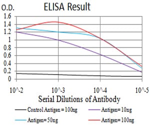
Figure 1: Black line: Control Antigen (100 ng);Purple line: Antigen (10ng); Blue line: Antigen (50 ng); Red line:Antigen (100 ng)
Western Blot
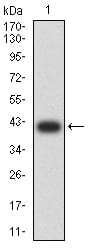
Figure 2:Western blot analysis using IL1R1 mAb against human IL1R1 (AA: 18-167) recombinant protein. (Expected MW is 40.8 kDa)
Western Blot
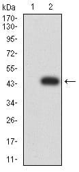
Figure 3:Western blot analysis using IL1R1 mAb against HEK293 (1) and IL1R1 (AA: 18-167)-hIgGFc transfected HEK293 (2) cell lysate.
Western Blot
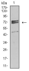
Figure 4:Western blot analysis using IL1R1 mouse mAb against Hela (1) cell lysate.
Flow cytometric
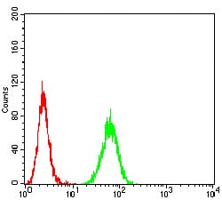
Figure 5:Flow cytometric analysis of Hela cells using IL1R1 mouse mAb (green) and negative control (red).
For Research Use Only. Not for use in diagnostic procedures.