

IghA1 Primary Antibody
Item Information
Catalog #
Size
Price
Description
IGHA1 (Immunoglobulin Heavy Constant Alpha 1) is a Protein Coding gene. Diseases associated with IGHA1 include pseudotumor cerebri. Among its related pathways are Vesicle-mediated transport and Regulation of nuclear SMAD2/3 signaling. GO annotations related to this gene include antigen binding and immunoglobulin receptor binding. An important paralog of this gene is IGHG4.
Product Overview
Entrez GenelD
3493
Aliases
IgA1
Clone#
7D12C5
Host / Isotype
Mouse / IgG1
Species Reactivity
Human, Mouse, Monkey
Immunogen
Purified recombinant fragment of human IghA1 (AA: 207-353) expressed in E. Coli.
Formulation
Purified antibody in PBS with 0.05% sodium azide
Storage
Store at 4°C short term. Aliquot and store at -20°C long term. Avoid freeze/thaw cycles.
Product Applications
WB (Western Blot)
1/500 - 1/2000
IHC_P(Immunohistochemistry)
1/200 - 1/1000
FCM (Flow Cytometry)
1/200 - 1/400
ELISA
1/10000
References
1.BMC Nephrol. 2014 Jun 13;15:89.
2.PLoS One. 2014 Feb 21;9(2):e89707.
2.PLoS One. 2014 Feb 21;9(2):e89707.
Product Image
Elisa
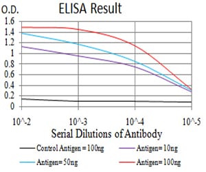
Figure 1: Black line: Control Antigen (100 ng);Purple line: Antigen (10ng); Blue line: Antigen (50 ng); Red line:Antigen (100 ng)
Western Blot
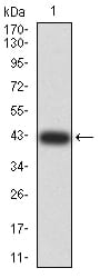
Figure 2:Western blot analysis using IghA1 mAb against human IghA1 (AA: 207-353) recombinant protein. (Expected MW is 41.7 kDa)
Western Blot
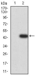
Figure 3:Western blot analysis using IghA1 mAb against HEK293 (1) and IghA1 (AA: 207-353)-hIgGFc transfected HEK293 (2) cell lysate.
Western Blot
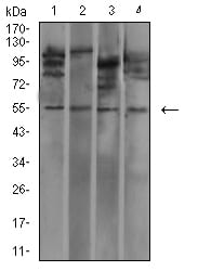
Figure 4:Western blot analysis using IghA1 mouse mAb against MOLT4 (1), L1210 (2), HepG2 (3), and COS7 (4) cell lysate.
Flow cytometric
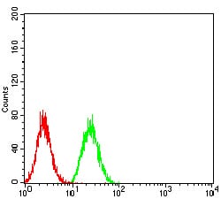
Figure 5:Flow cytometric analysis of Hela cells using IghA1 mouse mAb (green) and negative control (red).
Immunohistochemical analysis
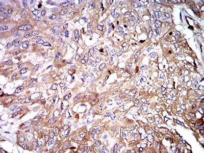
Figure 6:Immunohistochemical analysis of paraffin-embedded esophageal cancer tissues using IghA1 mouse mAb with DAB staining.
For Research Use Only. Not for use in diagnostic procedures.