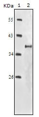
IGF1R-Beta Primary Antibody

Item Information
Catalog #
Size
Price
Description

IGF1R (insulin-like growth factor 1 receptor), a transmembrane receptor tyrosine kinase, is widely expressed in many cell types within fetal and postnatal tissues, and in many cell lines. Upon binding to its ligands, IGF-I and IGF-II, receptor autophosphorylation occurs. The triple tyrosine cluster within the kinase domain (Tyr1131, Tyr1135 and Tyr1136) is the earliest major site of autophosphorylation. Phosphorylation of these three tyrosine residues is necessary for kinase activation.Insulin receptors (IRs) share significant similarity with IGF1 receptors in both structure and function,including an equivalent triple tyrosine cluster within the activation loop of the kinase domain (Tyr1146, Tyr1150 and Tyr1151).Tyrosine autophosphorylation of insulin receptor is one of the earliest cellular responses to insulin stimulation. Autophosphorylation begins with phosphorylation of Tyr1146 and either Tyr1150 or Tyr1151. Full kinase activation requires the triple tyrosine phosphorylation.

Product Overview
Entrez GenelD
3480
Aliases
IGF1R, IGF1R-Beta
Clone#
3C8B1
Host / Isotype
Mouse / IgG2b
Species Reactivity
Human
Immunogen
Purified recombinant fragment of IGF1R-Beta expressed in E. Coli.
Formulation
Ascitic fluid containing 0.03% sodium azide.
Storage
4°C; -20°C for long term storage
Product Applications
WB (Western Blot)
1/500 - 1/2000
IHC_P(Immunohistochemistry)
1/200 - 1/1000
ELISA
1/10000
References
1. Adams, T.E. et al. Cell. Mol. Life Sci. 2000 57, 1050-1093.
2. Baserga, R. et al. Oncogene 2000 19, 5574-5581.
3. Scheidegger, K.J. et al. J. Biol. Chem. 2000 275, 38921-38928.
Product Image
Western Blot
Figure 1: Western blot analysis using IGF1R-Beta mouse mAb against truncated IGF1R-Beta recombinant protein.
Immunohistochemical analysis
Figure 2: Immunohistochemical analysis of paraffin-embedded human gastric adenocarcinoma(A), colon adenocarcinoma(B), endometrial carcinoma(uterus)(C), ovary adenocarcinoma(D), lung squamous cell carcinoma(E), stomach epithelium mucosae(F), showing membrane localization using IGF1R-Beta mouse mAb with DAB staining.
For Research Use Only. Not for use in diagnostic procedures.