
IFNAR1 Primary Antibody
Item Information
Catalog #
Size
Price
Description
The protein encoded by this gene is a type I membrane protein that forms one of the two chains of a receptor for interferons alpha and beta. Binding and activation of the receptor stimulates Janus protein kinases, which in turn phosphorylate several proteins, including STAT1 and STAT2. The protein belongs to the type II cytokine receptor family and functions as an antiviral factor.
Product Overview
Entrez GenelD
3454
Aliases
AVP; IFRC; IFNAR; IFNBR; IFN-alpha-REC
Clone#
2A5B12
Host / Isotype
Mouse / Mouse IgG1
Species Reactivity
Human, Mouse
Immunogen
Purified recombinant fragment of human IFNAR1 (AA: 28-156) expressed in E. Coli.
Formulation
Purified antibody in PBS with 0.05% sodium azide
Storage
Store at 4°C short term. Aliquot and store at -20°C long term. Avoid freeze/thaw cycles.
Product Applications
WB (Western Blot)
1/500 - 1/2000
IHC_P(Immunohistochemistry)
1/200 - 1/1000
FCM (Flow Cytometry)
1/200 - 1/400
ELISA
1/10000
References
1.J Clin Invest. 2021 Jan 4;131(1):e139980.
2.Front Immunol. 2021 Mar 5;12:628364.
2.Front Immunol. 2021 Mar 5;12:628364.
Product Image
Elisa

Figure 1:Black line: Control Antigen (100 ng);Purple line: Antigen (10ng); Blue line: Antigen (50 ng); Red line:Antigen (100 ng)
Western Blot

Figure 2:Western blot analysis using IFNAR1 mAb against human IFNAR1 (AA: 28-156) recombinant protein. (Expected MW is 40.8 kDa)
Western Blot

Figure 3:Western blot analysis using IFNAR1 mAb against HEK293-6e (1) and IFNAR1 (AA: 28-156)-hIgGFc transfected HEK293-6e (2) cell lysate.
Western Blot
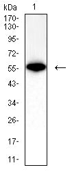
Figure 4:Western blot analysis using IFNAR1 mouse mAb against mouse lung (1) lysate.
Immunofluorescence analysis

Figure 5:Flow cytometric analysis of K562 cells using IFNAR1 mouse mAb (green) and negative control (red).
Immunohistochemical analysis

Figure 6:Immunohistochemical analysis of paraffin-embedded kidney cancer tissues using IFNAR1 mouse mAb with DAB staining.
Immunohistochemical analysis

Figure 7:Immunohistochemical analysis of paraffin-embedded rectum cancer tissues using IFNAR1 mouse mAb with DAB staining.
For Research Use Only. Not for use in diagnostic procedures.