

ID2 Primary Antibody
Item Information
Catalog #
Size
Price
Description
The protein encoded by this gene belongs to the inhibitor of DNA binding family, members of which are transcriptional regulators that contain a helix-loop-helix (HLH) domain but not a basic domain. Members of the inhibitor of DNA binding family inhibit the functions of basic helix-loop-helix transcription factors in a dominant-negative manner by suppressing their heterodimerization partners through the HLH domains. This protein may play a role in negatively regulating cell differentiation. A pseudogene of this gene is located on chromosome 3.
Product Overview
Entrez GenelD
3398
Aliases
GIG8; ID2A; ID2H; bHLHb26
Clone#
4E12G5
Host / Isotype
Mouse / IgG1
Species Reactivity
Human
Immunogen
Purified recombinant fragment of human ID2 (AA: 1-134) expressed in E. Coli.
Formulation
Ascitic fluid containing 0.03% sodium azide.
Storage
Store at 4°C short term. Aliquot and store at -20°C long term. Avoid freeze/thaw cycles.
Product Applications
WB (Western Blot)
1/500 - 1/2000
IHC_P(Immunohistochemistry)
1/200 - 1/1000
FCM (Flow Cytometry)
1/200 - 1/400
ELISA
1/10000
References
1.J Neurosci Res. 2012 May;90(5):925-32.
2.Mol Cancer. 2010 Jun 17;9:151.
2.Mol Cancer. 2010 Jun 17;9:151.
Product Image
Western Blot

Figure 1: Western blot analysis using ID2 mAb against human ID2 recombinant protein. (Expected MW is 17.3 kDa)
Flow cytometric

Figure 2: Flow cytometric analysis of SK-N-SH cells using ID2 mouse mAb (green) and negative control (purple).
Immunohistochemical analysis

Figure 3: Immunohistochemical analysis of paraffin-embedded breast cancer tissues using ID2 mouse mAb with DAB staining.
Immunohistochemical analysis

Figure 4: Immunohistochemical analysis of paraffin-embedded rectum cancer tissues using ID2 mouse mAb with DAB staining.
Elisa
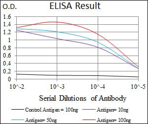
Black line: Control Antigen (100 ng); Purple line: Antigen(10ng); Blue line: Antigen (50 ng); Red line: Antigen (100 ng);
For Research Use Only. Not for use in diagnostic procedures.